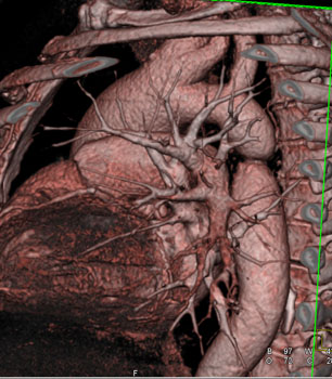
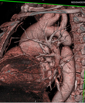
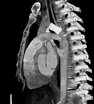
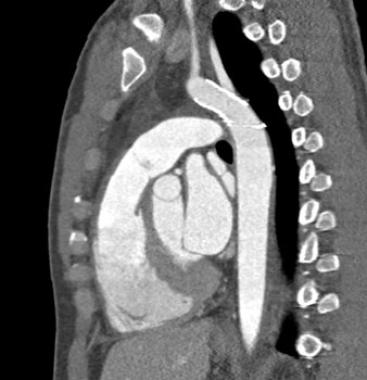
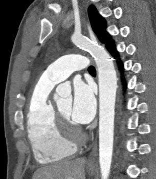
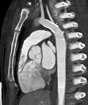
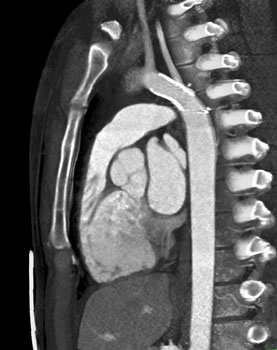
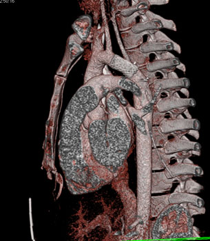
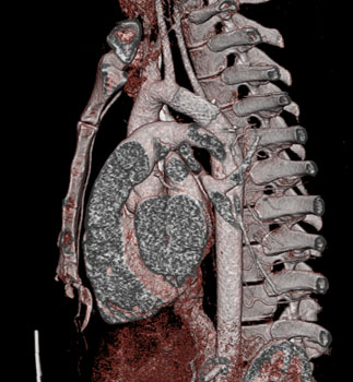
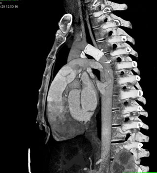
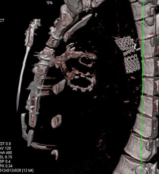
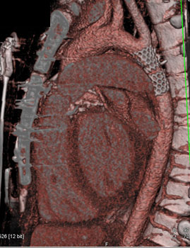
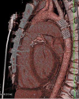
Diagnosis: Coarctation of the Aorta with Bicuspid Valve Coarctation of the Aorta: Facts - Aortic coarctation is narrowing of the aorta in the area where the ductus arteriosus inserts. Classically patients present with right arm hypertension and normal to low pressure in the legs. 3D and MPR Coronal CT imaging nicely depicts the focal aortic narrowing, with axial measurement contributing to full evaluation. Often there is post-stenotic aortic dilatation. In long standing or severe untreated coarctation, collaterals through the internal thoracic and subclavian arteries may develop.
- 5% of all congenital heart disease
- Narrowing of aorta most commonly just distal to the origin of the subclavian artery
- Simple coarctation means the absence of any other cardiac anomalies
- Complex aortic coarctation refers to associated anomalies plus COA
- Narrowing of aortic lumen results in LV hypertrophy
- Collaterals and rib notching are common
- May be isolated finding or associated with other cardiac issues
- Associated with cardiac anomalies
- Bicuspid valve (50%)
- VSD (33%)
- PDA (66%)
- Subaortic and mitral stenosis
- 20-30% of Turners syndrome patients have a COA
- Males > females by 2-1
Congenital Anomalies of the Thoracic Aorta - Sequestration
- Vascular rings
- Coarctation of the aorta with dilated ascending aorta
- Aberrant vessels
- Right sided arch and associated anomalies
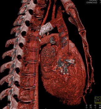
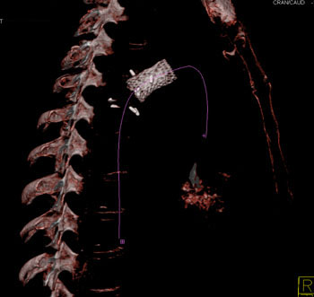
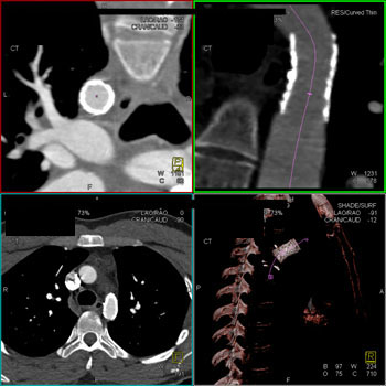
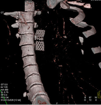
CTA s/p COA Repair After repair of the coarctation with graft CT is excellent at defining graft patency and flow. Use of 3D and vessel analysis pool in this regard is critical as the following images nicely demonstrate. Successful Stent Repair of COA |