Cinematic Rendering: Principles, Applications, and Observations in CTCinematic Rendering: Principles, Applications, and Observations in CT Steven P. Rowe, M.D., Ph.D. Elliot K. Fishman, M.D. The Russell H. Morgan Department of Radiology and Radiological Science Johns Hopkins University School of Medicine Baltimore, MD, USA |
Cinematic Rendering (CR)
Johnson PT, Schneider R, Lugo-Fagundo C, et al. AJR Am J Roentgenol. 2017;209:309-312. |
Cinematic Vs. Volume Rendering
|
Cinematic Vs. Volume Rendering
|
Cinematic Rendering: Strikingly Different from other 3D Methods CR uses a global lighting model that creates effects including realistic shadowing, appropriate obscuration of the light source depending on the relationship with the light source and the image in the volume, etc. Johnson PT, Schneider R, Lugo-Fagundo C, et al. AJR Am J Roentgenol. 2017;209:309-312. |
Clinical Utility of Cinematic Rendering has not Been Established
|
Clinical Examples of Cinematic Rendering
|
Calvarial, Skull Base, and Maxillofacial Normal Anatomy 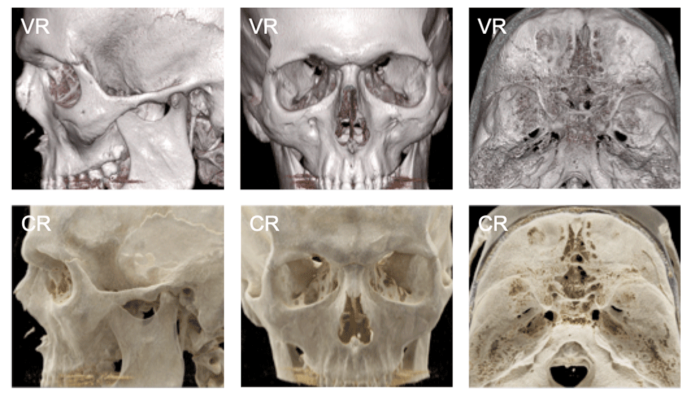 VR vs. CR: normal skull anatomy |
Calvarial, Skull Base, and Maxillofacial Normal Anatomy 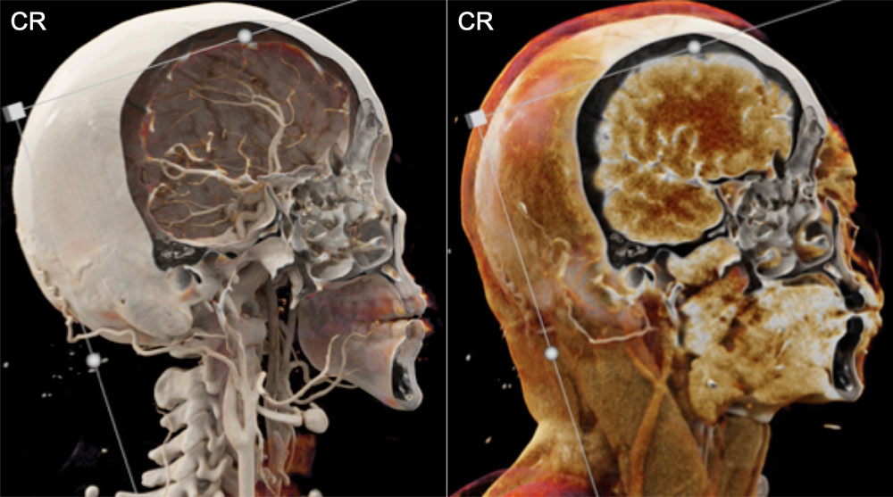 |
Calvarial, Skull Base, and Maxillofacial Normal Anatomy 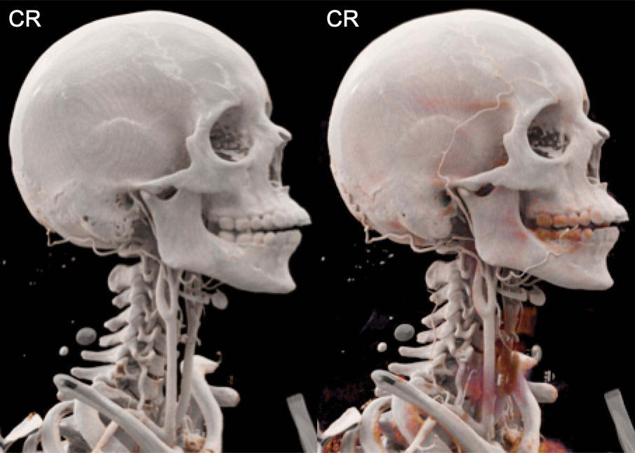 Photorealistic images such as these cinematic renders may prove invaluable in trainee education |
Calvarial, Skull Base, and Maxillofacial Normal Anatomy 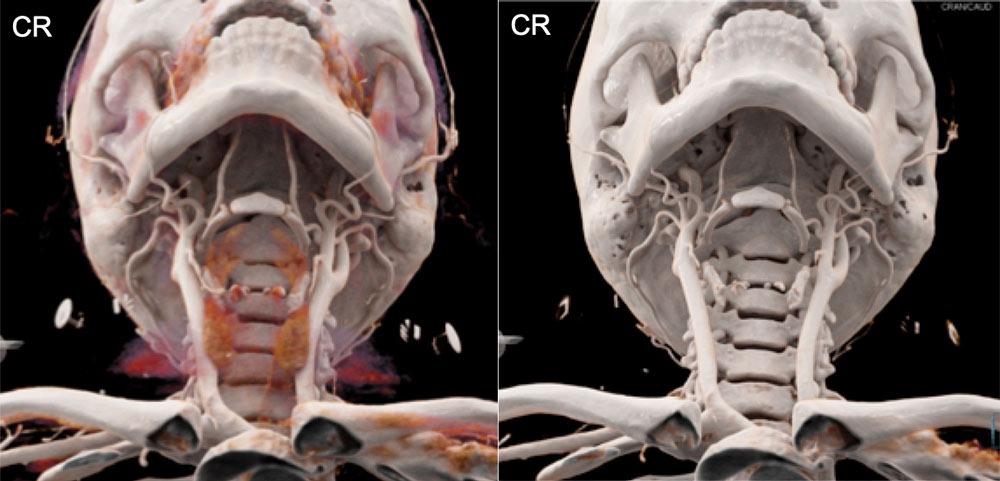 |
Forearm and Hand Anatomy with Varying Window Widths and Levels As with any 3D technique, an important aspect to the creation of diagnostically useful images is the real-time interaction of the radiologist with the software to ensure that all relevant pathology is appropriately displayed. |
Cinematic Rendering of Forearm and Hand 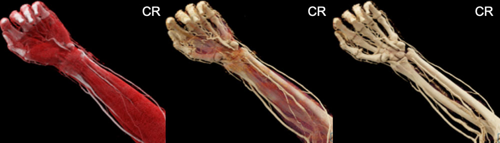 The potential for CR to replace MRI for soft tissue and tendon evaluation in certain situations should be extensively investigated |
Cinematic Rendering of Forearm and Hand 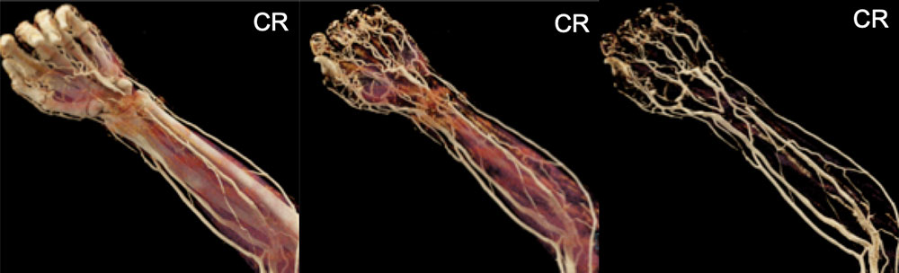 Changes in widow width, window setting, and transparency can allow for almost any tissue or combination of tissues to be displayed in the visualizations |
Cinematic Rendering of Forearm and Hand 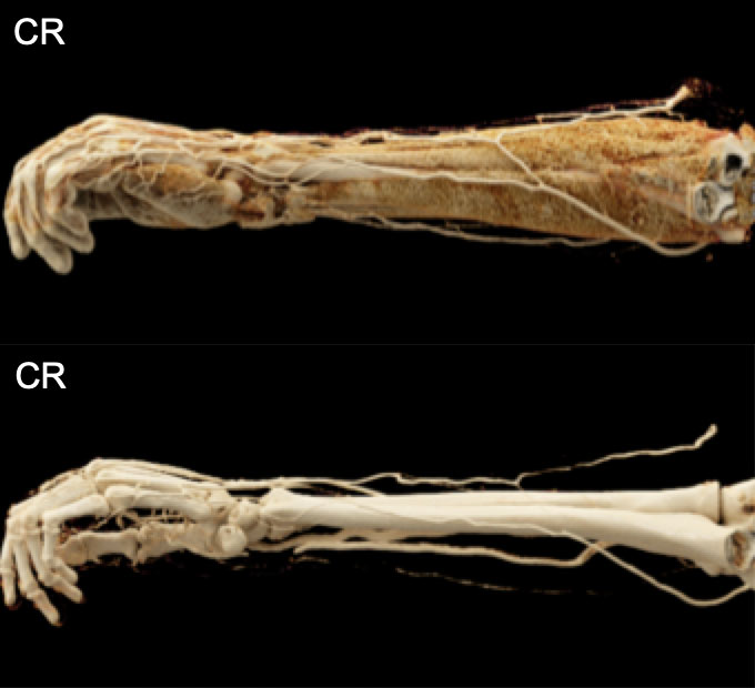 Viewing the volume from different virtual points of view will ensure that key findings are not missed because of the realistic shadowing effects/obscuration of the light source that are intrinsic to the method |
Normal Fetal and Placental Anatomy can be Well-Evaluated Note the shadowing that occurs between the pubic symphysis and the fetal skull in the last two images (red arrowheads) that allows for a definitive understanding of the relative locations of those objects 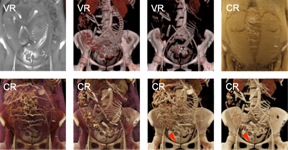 Rowe SP, Fishman EK. Radiol Case Rep. 2017; Ahead of print. |
Highly Detailed Head and Neck Soft Tissue Anatomy can be Rendered 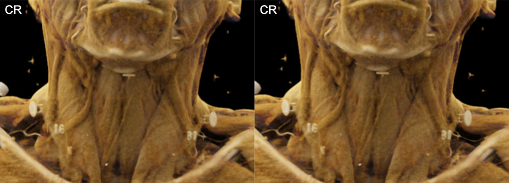 |
Highly Detailed Head and Neck Soft Tissue Anatomy can be Rendered 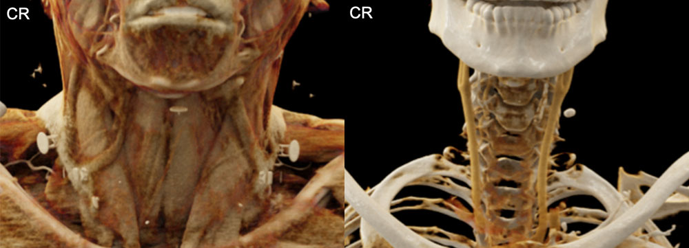 |
Highly Detailed Head and Neck Soft Tissue Anatomy can be Rendered 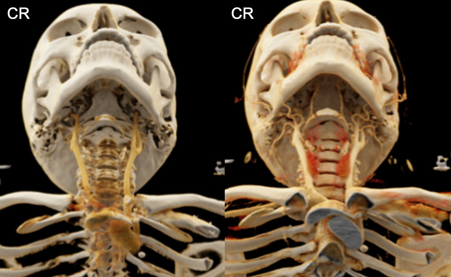 |
Highly Detailed Head and Neck Soft Tissue Anatomy can be Rendered 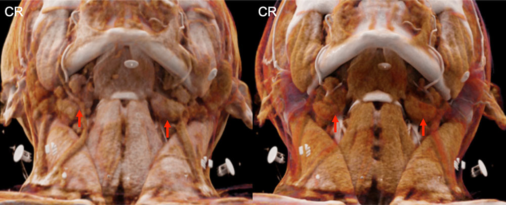 Small structures, such as the submandibular glands (red arrows) are delineated on CR images, and aspects of the textures of the glands can be appreciated. |
Normal Stomach Anatomy 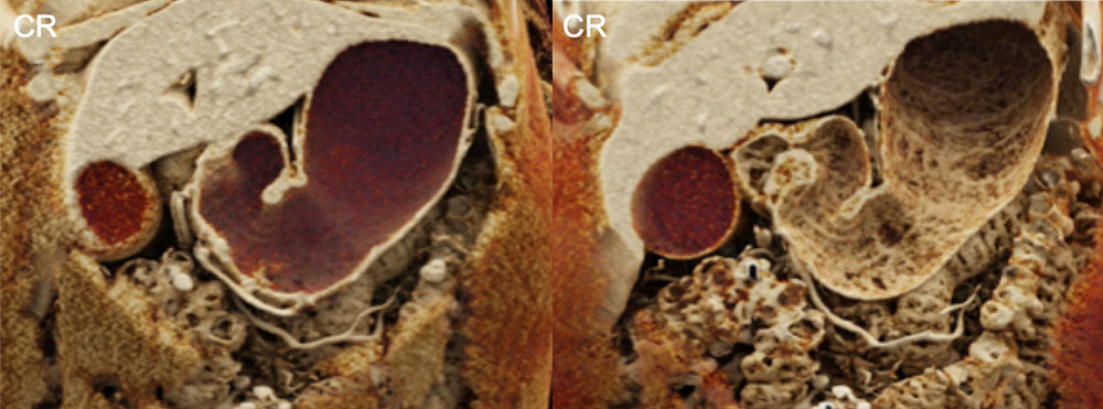 The level of surface detail that can be achieved with CR allows the texture of the folds of the stomach to be depicted in the second of these panels |
Destructive Facial Squamous Cell Carcinoma 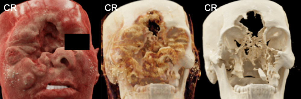 CR of facial squamous cell carcinoma – appropriate window settings allow one to “move through” the layers of soft tissue to display more of the underlying bone structure |
Squamous Cell Carcinoma Invading the Calvarium 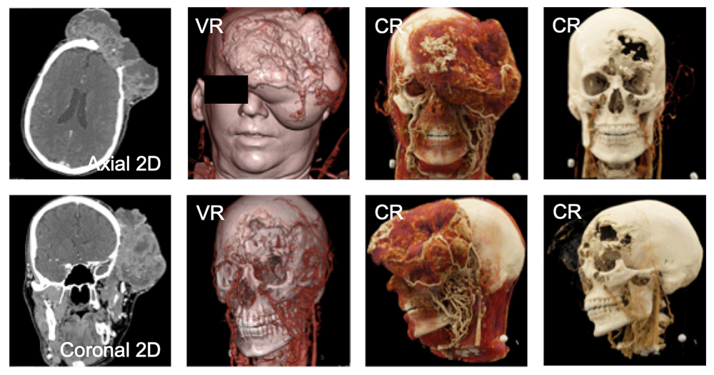 Note the excellent display of the vascular map of the lesion on the CR images as well as the photorealistic appearance of the body calvarium |
Calvarial, Skull Base, and Maxillofacial Anatomy and Pathology 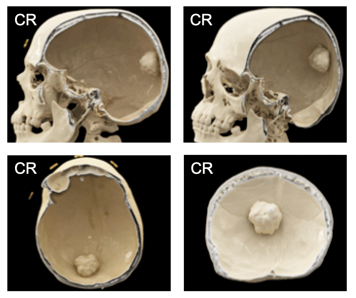 Posterior calvarial osteoma: note the shadowing around the interface between the osteoma and the overlying skull, indicating that this lesion arises from a bony stalk |
Cinematic Rendering of Pancreas Serous Cystadenoma 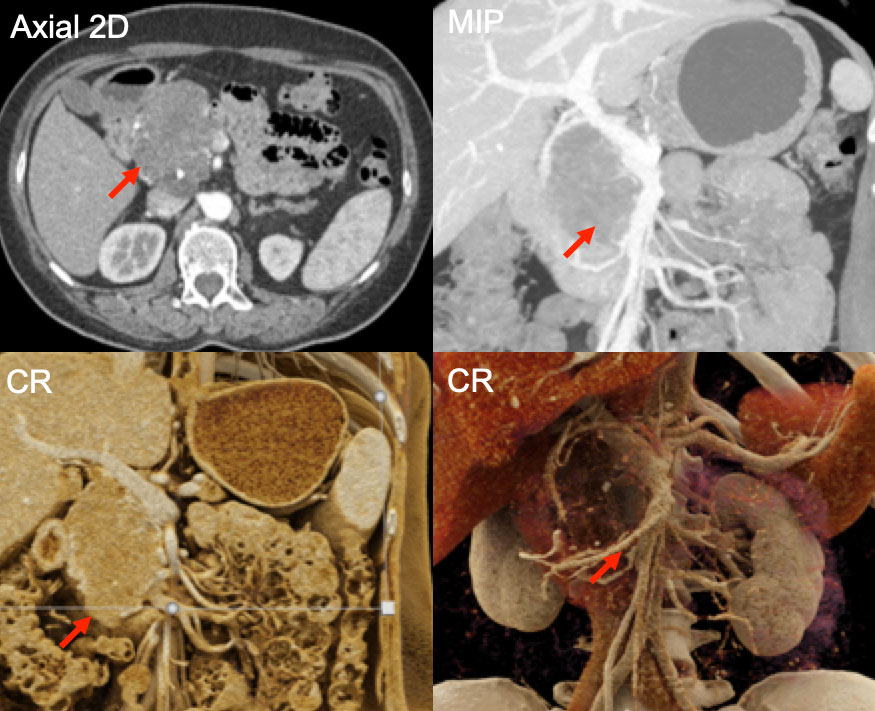 |
Pancreatic Lipoma 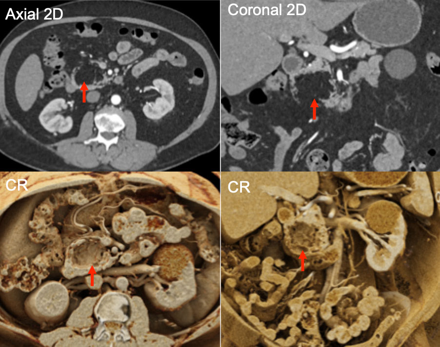 |
Adencarcinoma of the Tail of the Pancreas 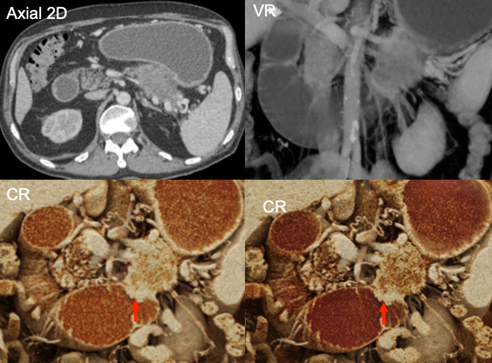 Note the excellent delineation of the duodenal invasaion (red arrows) on the CR images |
Pancreatic Adenocarcinoma with Vascular Invasion 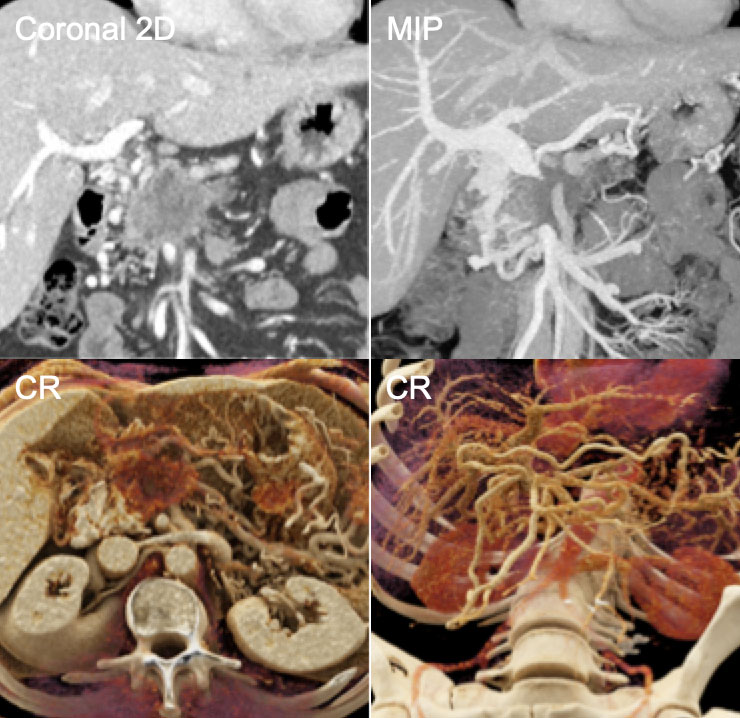 Note the ability of CR to display the abdominal vasculature including the collateral vessels in a photorealistic manner. |
Pancreatic Adenocarcinoma with Upstream Atrophy 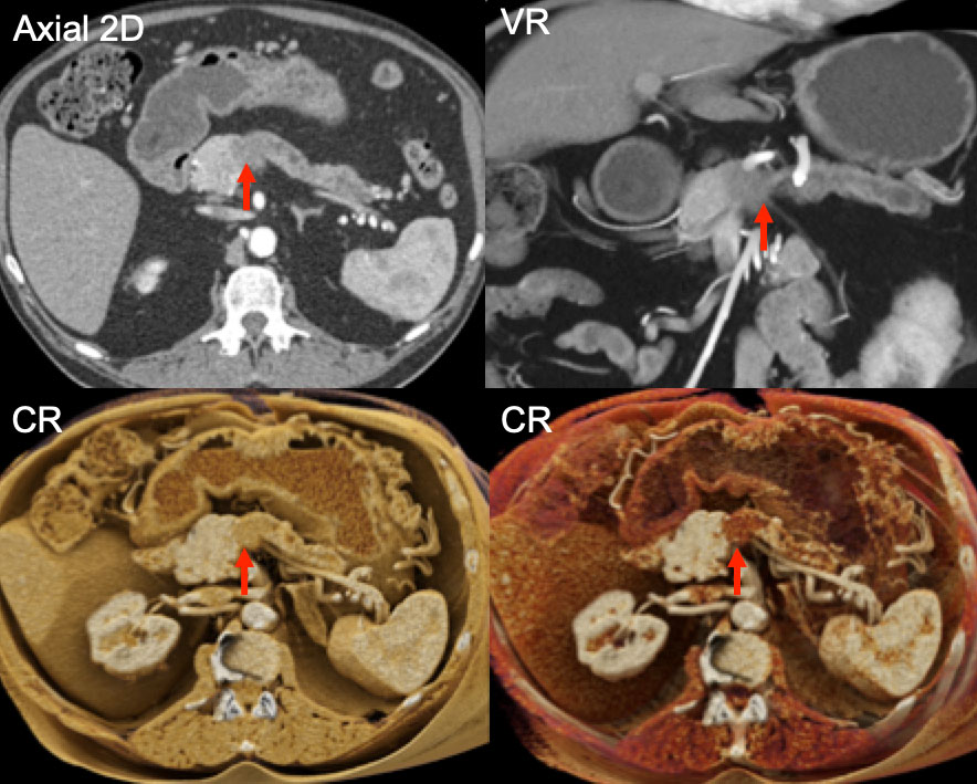 Note the texture change that is visible between normal pancreas, the tumor (red arrows) and the atrophied pancreatic tail on the CR images. |
Pancreatic Neuroendocrine Tumor with Extensive Neovascularity 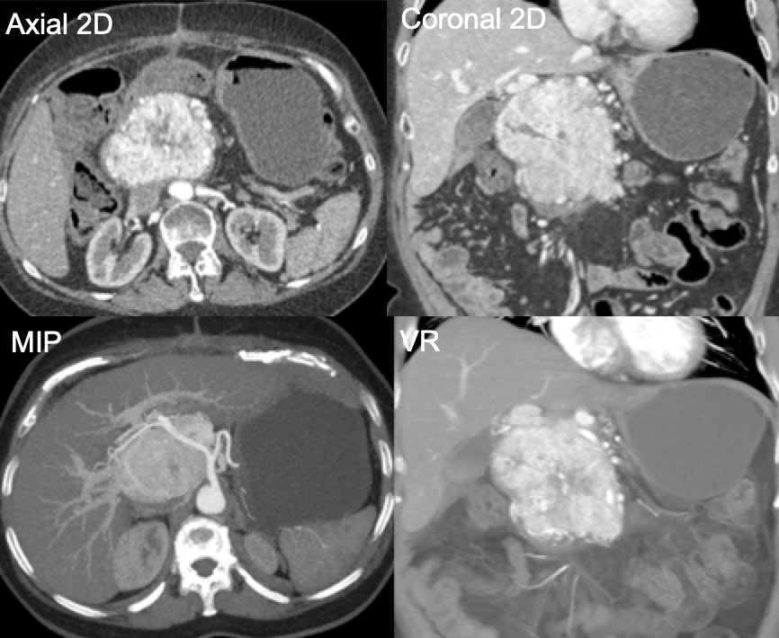 |
Pancreatic Neuroendocrine Tumor with Extensive Neovascularity 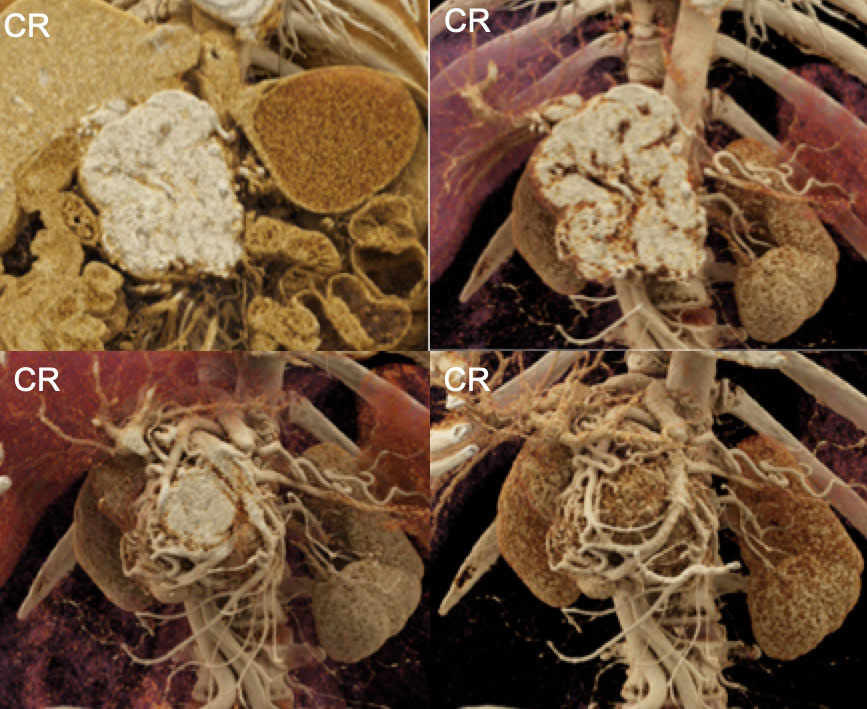 Note the effective depiction of the “nest” of tumor neovascularity that surrounds the tumor in the bottom panels. |
Large, Highly-Vascular Gastro-Intestinal Stromal Tumor 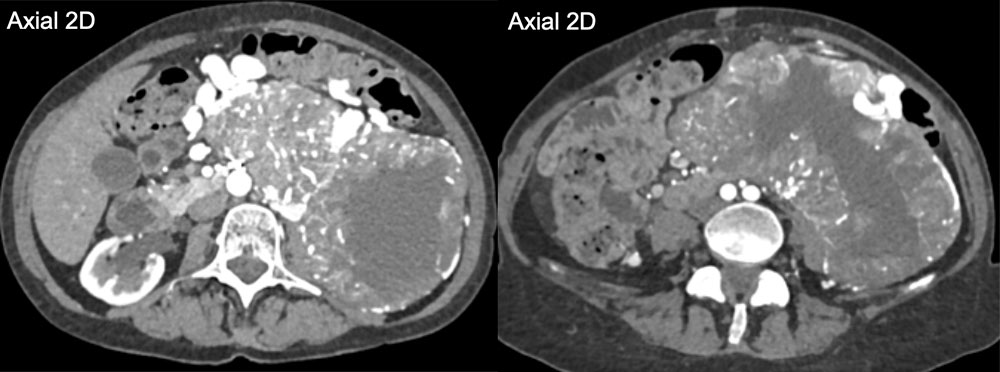 |
Large, Highly-Vascular Gastro-Intestinal Stromal Tumor 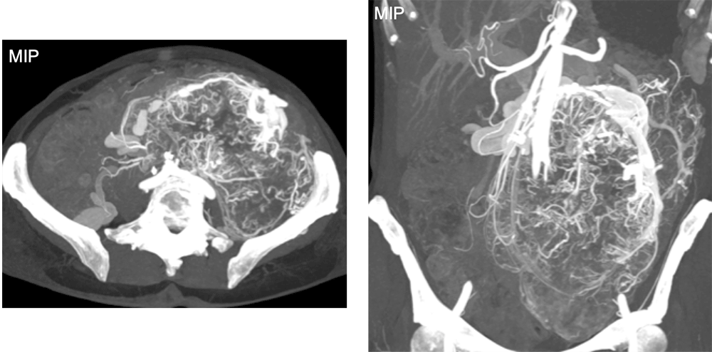 |
Large, Highly-Vascular Gastro-Intestinal Stromal Tumor 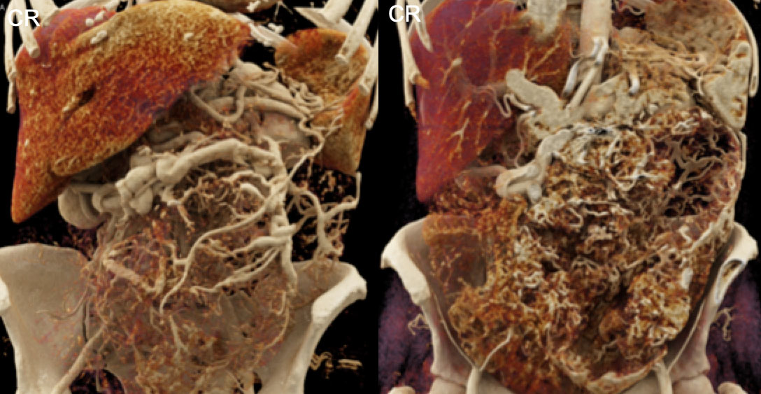 |
Large, Highly-Vascular Gastro-Intestinal Stromal Tumor 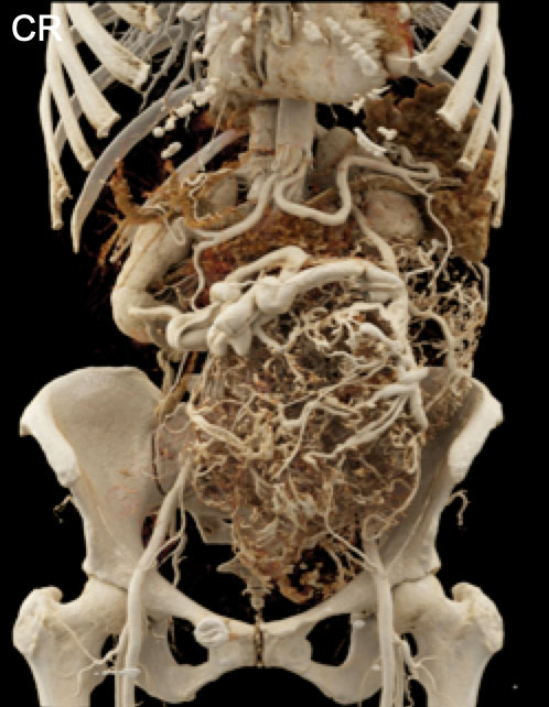 For a large, hyper-vascular tumor such as this GIST lesion, the combination of accurate vascular maps and high levels of soft tissue detail that can be provided by CR may prove very beneficial for pre-operative planning |
Right Lower Pole Clear Cell Renal Cell Carcinoma 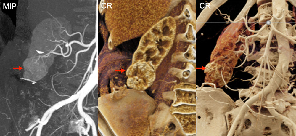 |
Left Lower Pole Clear Cell Renal Cell Carcinoma 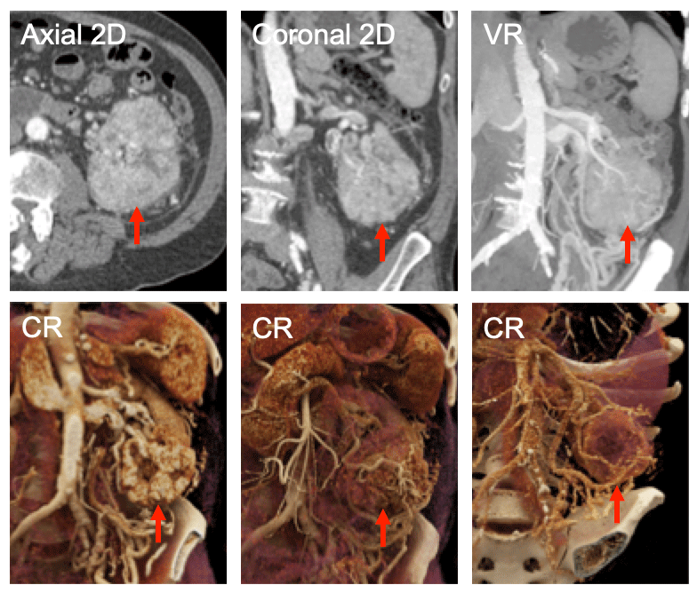 The texture of the tumor and the extensive neovascularity are shown to good advantage by the CR images (red arrows in the bottom row of figures. Pre-operative planning in such complex cases may prove to be a valuable application for this 3D visualization technique |
5 mm Clear Cell Renal Cell Carcinoma Detected Incidentally 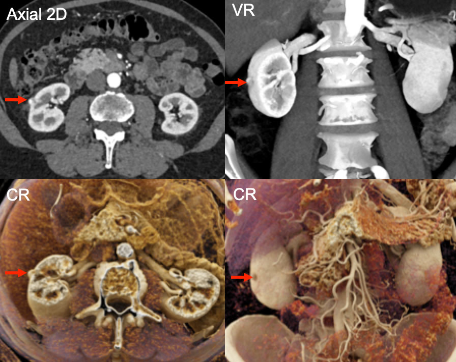 |
Infiltrating Right Kidney Renal Cell Carcinoma 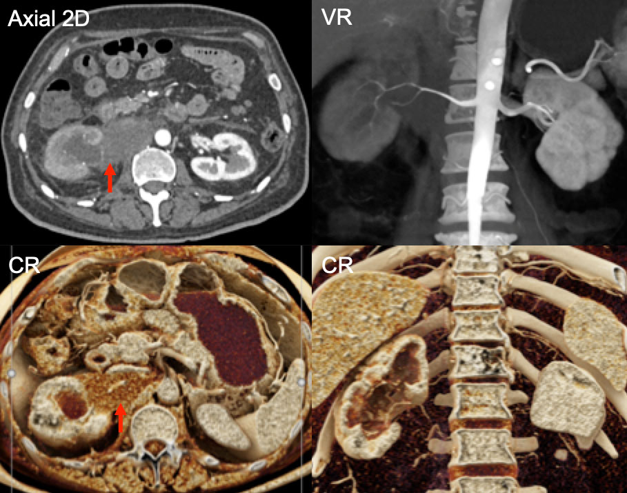 |
Left Kidney Transitional Cell Carcinoma 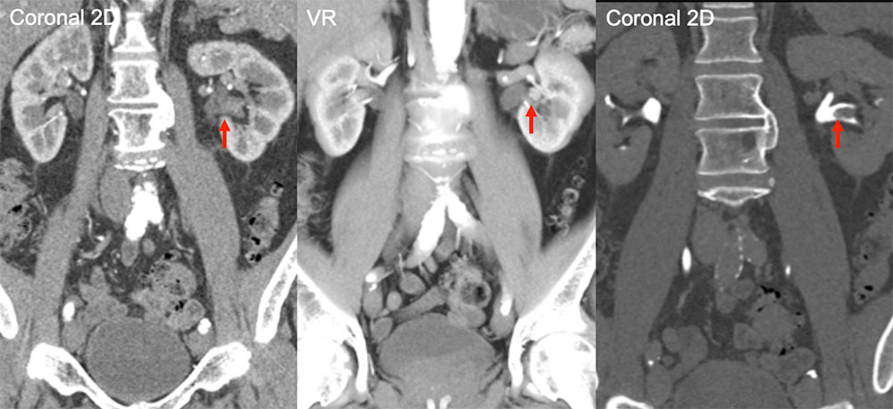 Careful scrutiny of the 2D and VR images demonstrates a lesion (red arrows) in the left renal collecting system. |
Left Kidney Transitional Cell Carcinoma 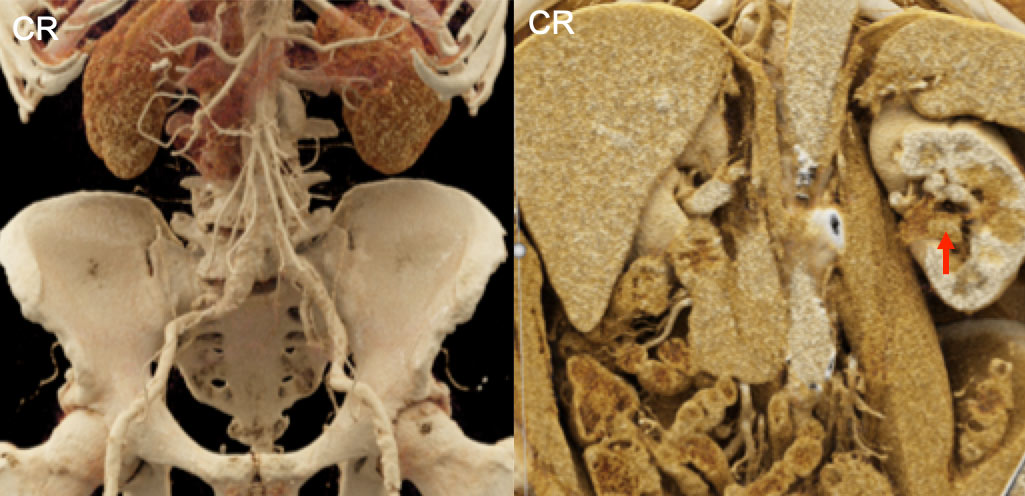 |
Left Kidney Transitional Cell Carcinoma 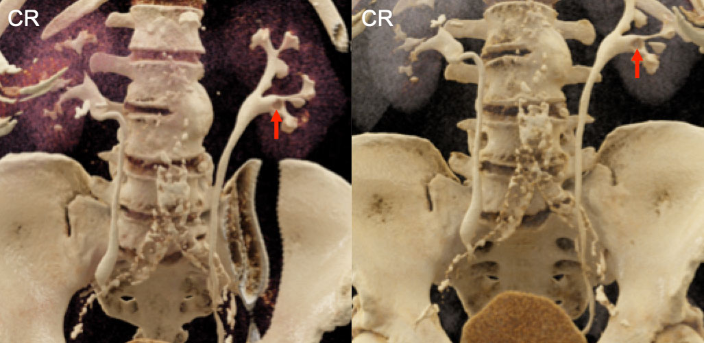 |
Another Example of Left Kidney Transitional Cell Carcinoma 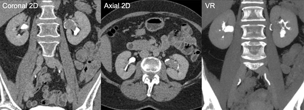 |
Another Example of Left Kidney Transitional Cell Carcinoma 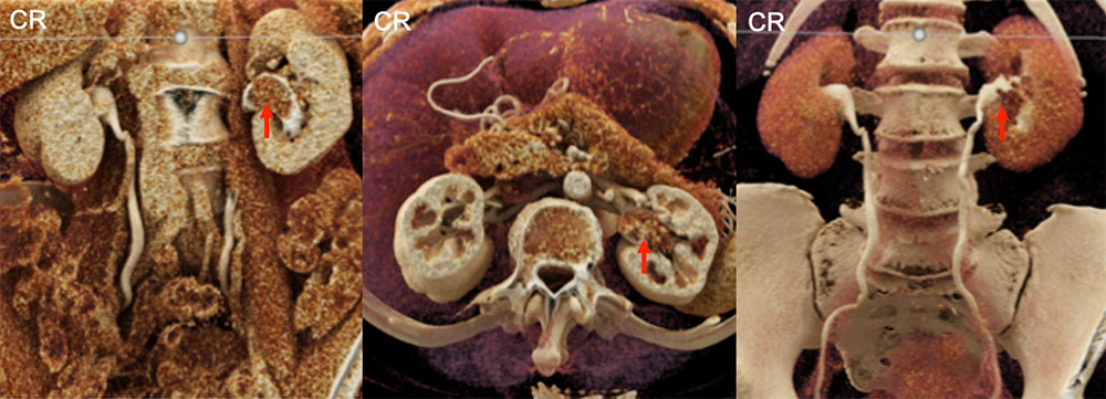 Depending on the contrast phase and the widow width and settings used, a transitional cell carcinoma (red arrows) can appear as either a mass lesion or a defect within an excreted contrast column |
Colorectal Metastasis to the Liver 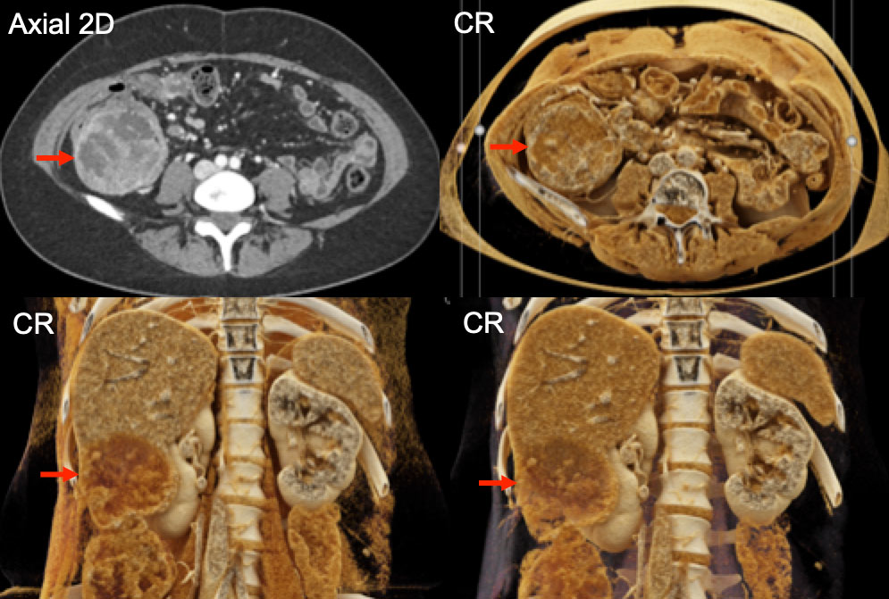 |
Renal Cell Carcinoma Adrenal Metastases 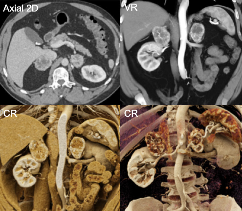 Although the 2D and VR images demonstrate heterogeneity in these renal cell carcinoma metastases, the texture of that heterogeneity is much more apparent in the CR panels on the bottom |
Metastatic Carcinoid Tumor 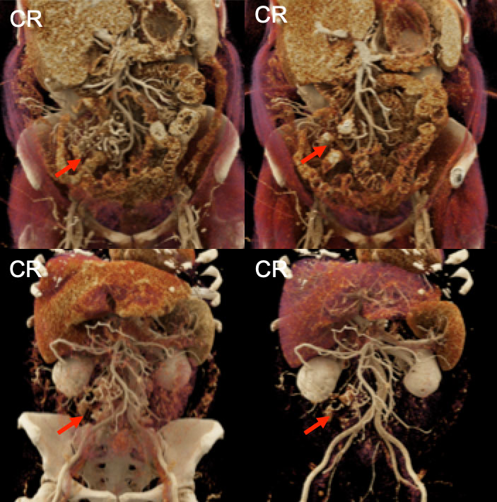 This example of metastatic carcinoid tumor (red arrows) to the mesentery has the typical calcifications and vascular tethering, with the effects on the vasculature well displayed on these CR images |
Small Bowel Gastrointestinal Stromal Tumor 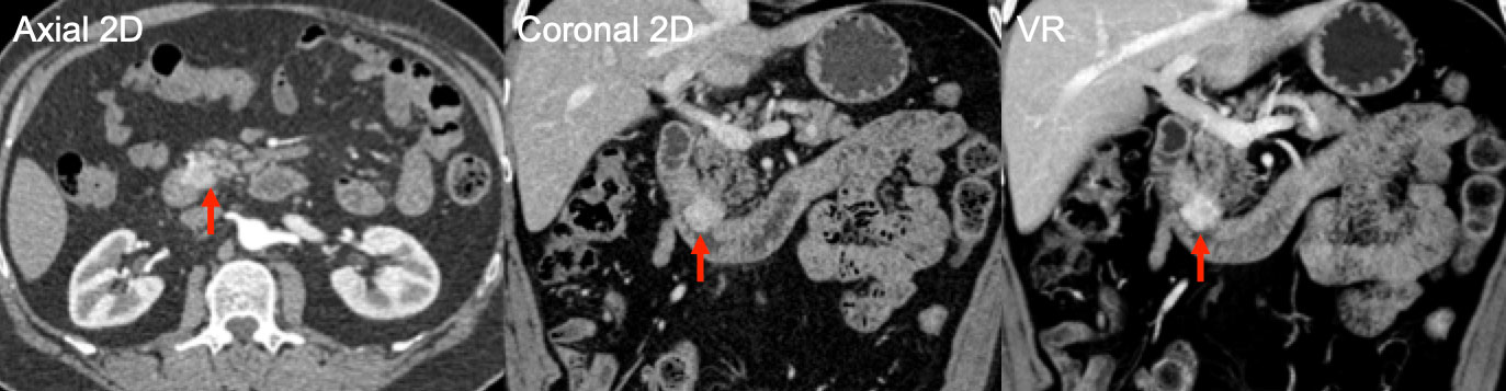 |
Small Bowel Gastrointestinal Stromal Tumor 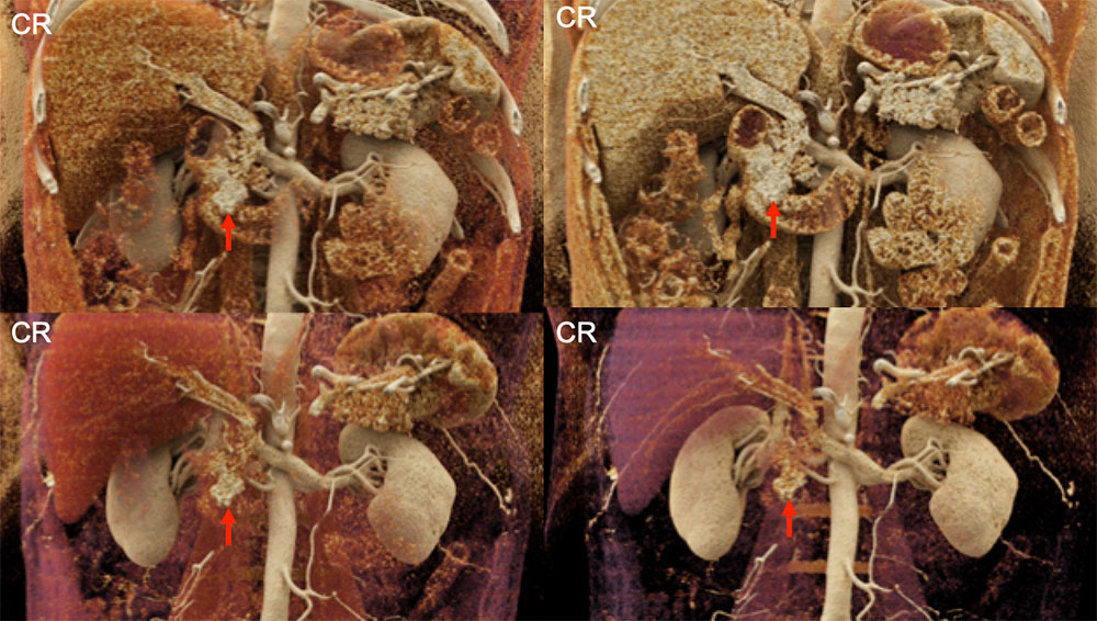 |
Small Bowel Obstruction Secondary to Cecal Adenocarcinoma 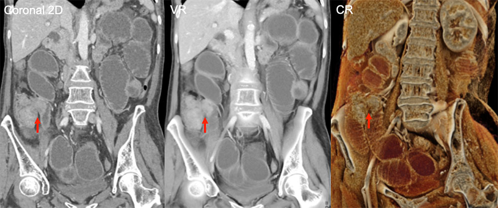 |
Treated Gastrointestinal Stromal Tumor Liver Metastasis 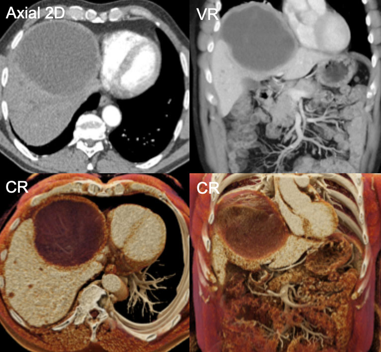 |
Liver Hemangioma 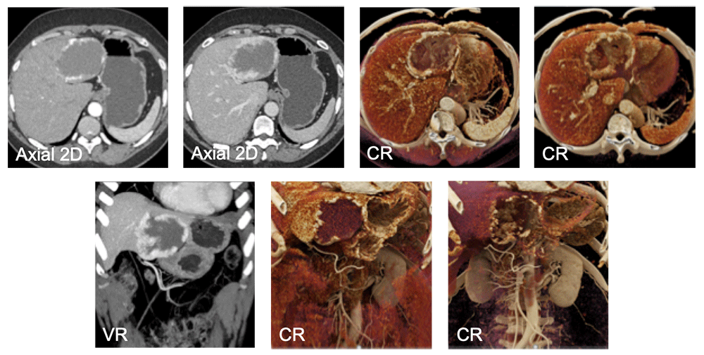 |
Carcinoid Liver Metastases 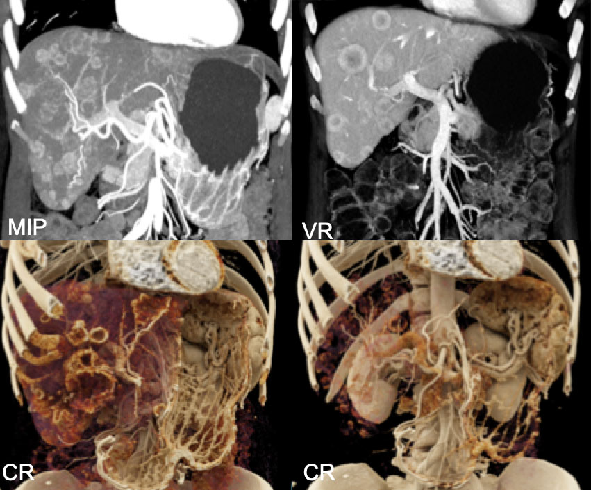 |
Gallbladder Adenocarcinoma 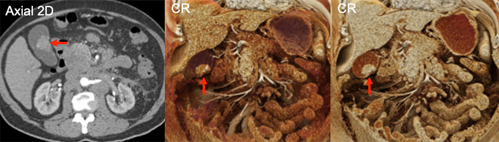 |
