Vascular Pathology in Loeys-Dietz Syndrome: The Role of 64 row MDCT with 3D Rendering for Vascular Screening and Detection of Vascular Abnormalities
Vascular Pathology in Loeys-Dietz Syndrome: The Role of 64 row MDCT with 3D Rendering for Vascular Screening and Detection of Vascular Abnormalities Russell H. Morgan Department of Radiology and Radiological Science, Johns Hopkins School of Medicine |
Loeys-Dietz Syndrome (LDS) is a recently identified genetic syndrome, with phenotypic resemblance to Marfan syndrome (MFS), Marfanoid craniosynostosis syndrome (Shprintzen-Goldberg syndrome) and in some cases, Ehlers-Danlos type IV (EDS-IV). |
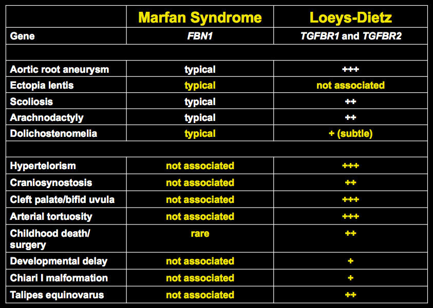
Characteristic Traits of LDS (common)
|
Characteristic Traits of LDS (less common)
|
Loeys Dietz Syndrome: Clinical Subtypes LDS type I:
|
Loeys Dietz Syndrome: Clinical Subtypes LDS type II:
|
Phenotypic spectrum and pathogenesis of a new aortic aneurysm syndrome caused by mutations in TGFBR1 and TGFBR2 |
 |
LDS: Thoracic CT Findings
|
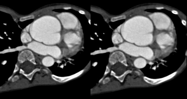
LDS: Aortic Root Aneurysm
|
Aortic Root Aneurysm
|
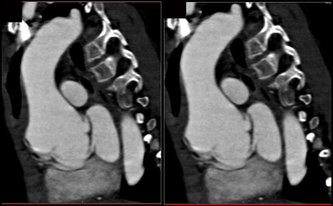 Dilated Aortic Root |
 |
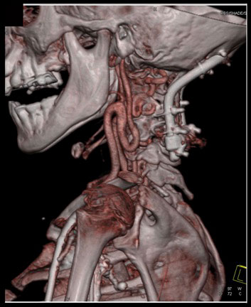
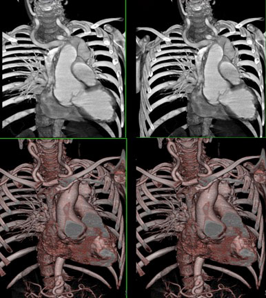 Dilated aortic root as well ectatic carotid arteries |
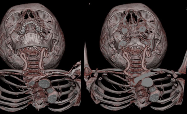
Carotid Arteries and Intracranial Vessels: CT Findings
|
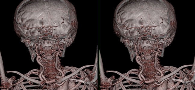 Tortuous Carotid Arteries |
 |
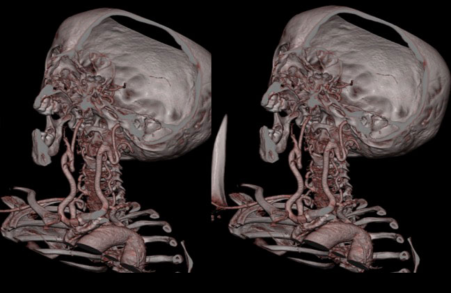 |
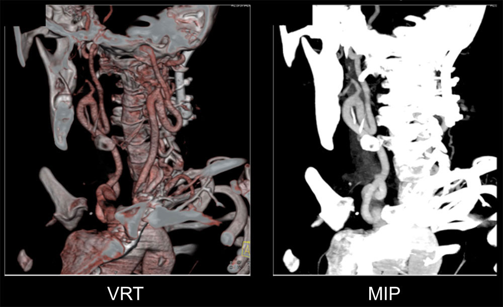 Loeys-Dietz Syndrome: Ectatic Carotid Artery |
|
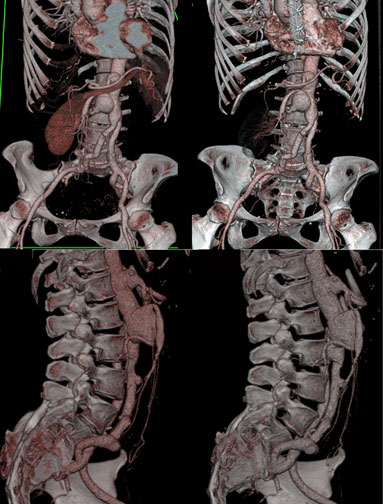
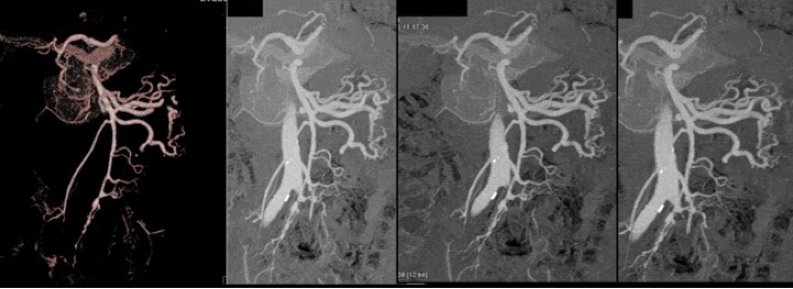
Abdominal Aorta: CT Findings
|
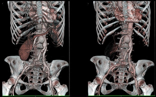 This case is unique with so many aneurysms seen off the abdominal aorta, as well as the origins of the celiac, SMA, IMA and smaller branch vessels. |
 |
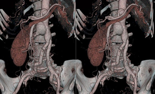 |
 Small Aneurysms of the SMA |
Conclusion
|