google ads

Short and Long Term Complications of Endovascular Stents: MDCT Findings and the Importance of Volumetric Visualization with 2D MPRs and 3D Rendering for Detection and Characterization

Short and Long Term Complications of Endovascular Stents: MDCT Findings and the Importance of Volumetric Visualization with 2D MPRs and 3D Rendering for Detection and Characterization

Elliot K. Fishman, MD, FACR


Type I Endoleak

  • Aneursymal blood flow through poorly apposed attachment sites
    • A: Proximal landing zone
    • B: Distal landing zone
    • C: Failure of iliac occluder
  • Occurs in 10% of EVAR procedures
  • Direct communication with arterial circulation
    • Best visualized on arterial phase imaging
    • Contrast in aneurysm sac is apposed to attachment zones
  • Chimney Grafts are predisposed to gutter leaks; however, many times spontaneously resolve
  • Very few Type I are self limiting, with most requiring treatment
    • Balloon Angioplasty
    • Stent-Graft Extension
    • Convert bifurcated devices into aorto-uniliac device with fem-fem bypass
    • Embolization


Type I Endoleak

Type I Endoleak


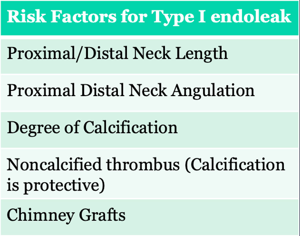
Type I Endoleak

60 year old woman with EVAR presented to ED with pain. Coronal IV CECT images show a new Type I endoleak (red arrows) and stent deformity (yellow arrows) in setting of infection (see slide 24)
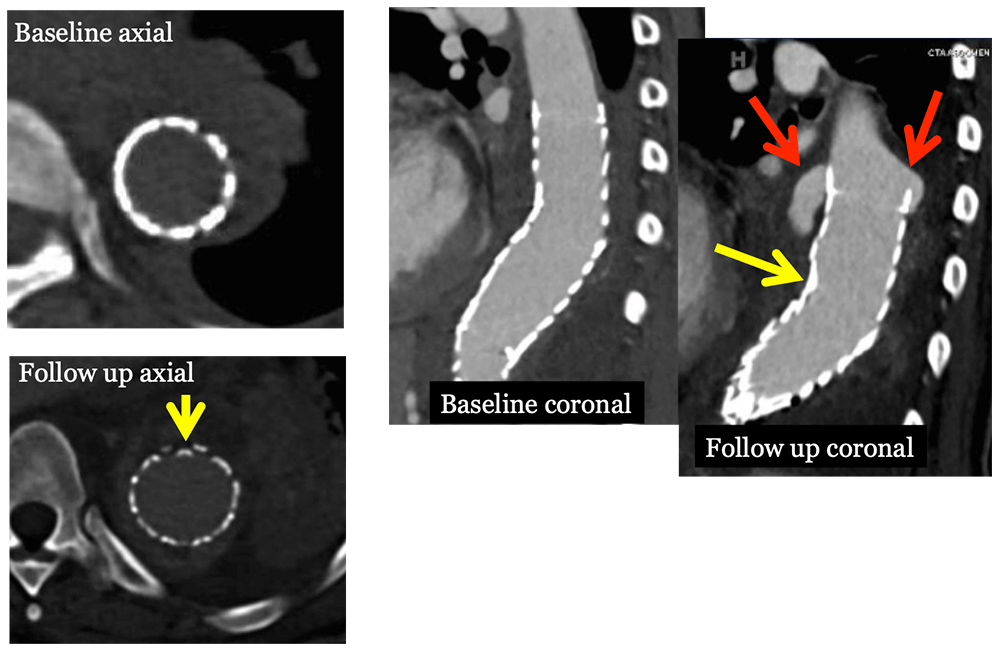
Type I Endoleak


Type II Endoleak

  • Aneurysmal blood flow retrograde from collateral vasculature
    • Inferior Mesenteric Artery – enters anteriorly
    • Lumbar Arteries – enters posterolateraly
    • Contrast appears peripherally in aneurysm sac
  • Most common type of endoleak, occurring in 10-25% of cases
  • Reportedly, 40-80% spontaneously resolve due to low pressure system within one year.
  • Delayed or persistent appearance after 1 year heralds sac growth.
  • Treatment remains controversial with most centers treating once sac growth exceeds 5 -10 mm.
    • Embolization
      • transarterial and translumbar
    • Laparoscopic vs Open approach
Type II Endoleak


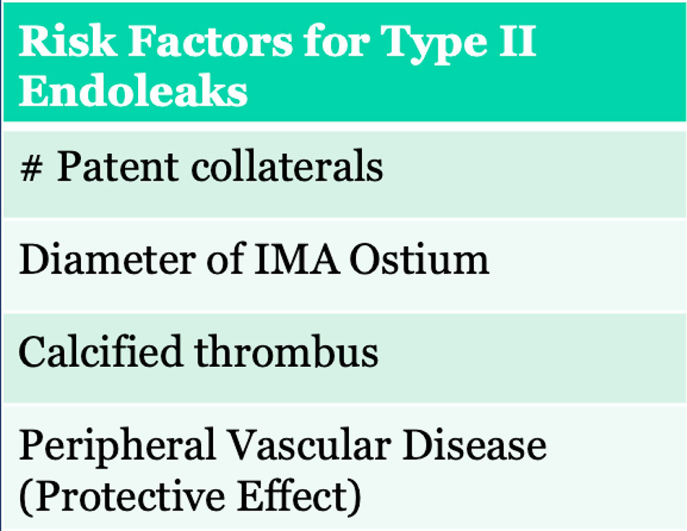
Type II Endoleak

Small type II endoleak (circle) in anterior sac. Search for feeding vessel, which is typically the inferior mesenteric artery (arrow) with anterior endoleaks.
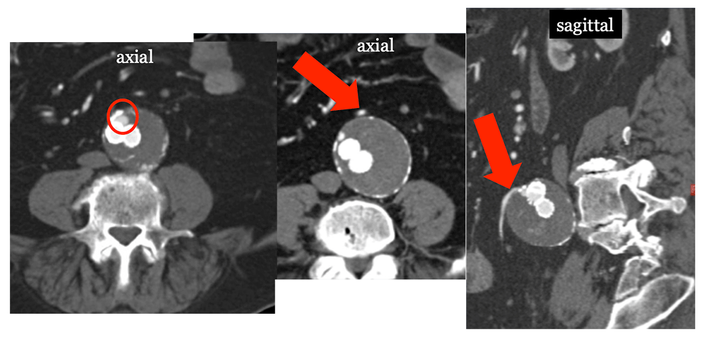
Type II Endoleak


Type III Endoleak

  • Aneurysmal blood flow through the body of the stent graft
    • Fabric tear, device fracture, dissociation
    • Device migration
    • Device kinking/occlusions may result in leak
  • High arterial pressures require emergent treatment, similar to type I
  • Contrast is seen adjacent to the body of the endograft
  • Subtle erosions of the graft may only be seen with multiplanar reformations
  • Repair depends on cause of leak
    • Additional stent/limb extension
    • Conversion to aortouniliac with bypass
    • PTA +/- stent
    • Thrombectomy +/- stent
    • Open conversion


Type III Endoleak

75 Year old male with history of EVAR presenting with back pain. MDCT shows kinked stent graft (yellow arrow) causing type III endoleak (circle), which resulted in rupture (red arrow).
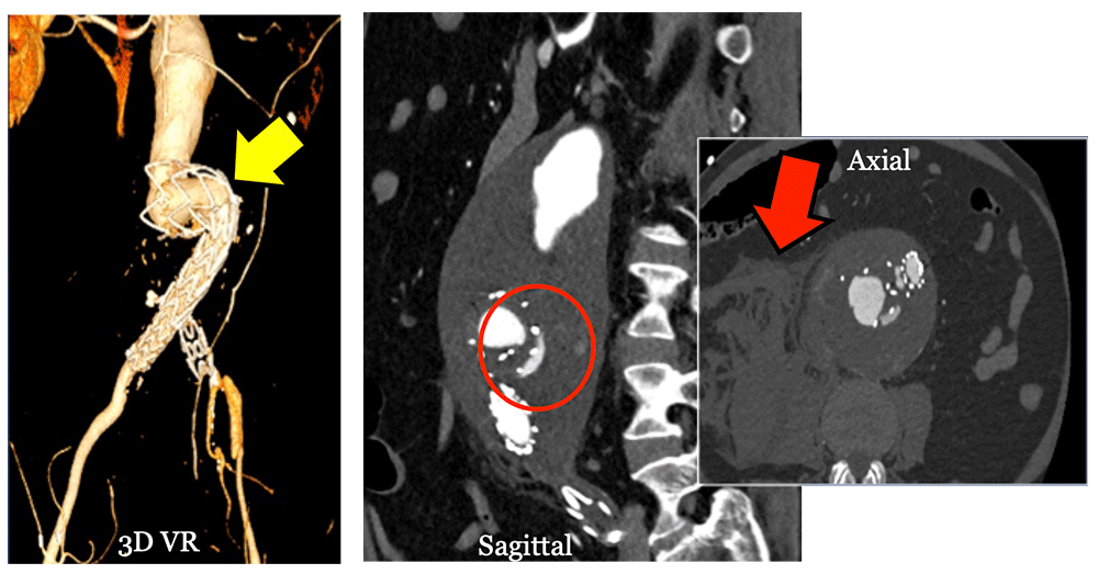
Type III Endoleak


Type IV Endoleak

  • Aneurysmal flow through stent-graft pores
  • Identified at implantation as “blush” on immediate post angiogram
  • Require no intervention


Type V Endoleak

  • Endotension is referred to as expansion of the aneurysm without presence of endoleak
  • May be related to occult type I, II, or III endoleak
  • Pressure transmission from the graft, infection or hygromas
  • May be present when aneurysm sacs do not shrink in size


Migration and Kinking

  • Migration leads to
    • Endoleak
    • Kinking
    • Rupture
  • Risk factors for migration
    • Duration
    • Degree of oversizing
    • Neck diameter
    • Length landing zone
  • Treatment
    • Stent graft extension
    • Open repair
  • Kinking leads to
    • Endoleak
    • Migration
    • Thrombosis
  • Risk Factors for kinking
    • Severe angulation
    • Migration
    • Narrow aortic bifurcation


Dissociation of components

77 year old male s/p EVAR for AAA. Routine IV CECT at 1 year follow up revealed stent graft dissociation (yellow arrows), aneurysm sac increase (circle) to 10 cm and large type III leak (E) in the absence of symptoms, probably sequela of recent fall.

Dissociation of components


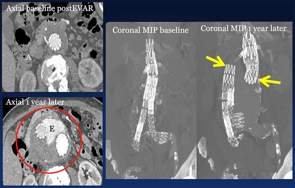
Thrombosis, Occlusion and Stenosis

  • Risk Factors
    • Migration, Kinking
    • Oversizing, Infolding/Twisting of graft material
    • Tortuous, diminutive iliac arteries
  • Typical treatment for stenosis and occlusion includes
    • Aortouniliac devices with fem-fem bypass
    • Thrombectomy or bypass for thrombosis
    • Ballon angioplasty +/- stenting for stenosis


Infection

  • Occurs in less than 1%, however associated with high mortality.
  • Sources include procedure contamination (perioperative) vs disseminated infection (late)
    • Kidney stone, appendicitis induced peritonitis, inflammatory aneurysm, pneumonia
  • Important to identify inflammation adjacent to the stent graft
    • Peri-graft fluid collections
    • Abnormal enhancement
    • Gas bubbles
  • Aneurysm expansion may lead to erosion into bowel, fistulization and infection
  • Requires surgical repair


Infected EVAR and Type I Endoleak

60 year old woman with EVAR presented to ED with pain (same patient as slide 14). Coronal IV CECT images show a new Type I endoleak (red arrows) in the setting of lung infection. Note infected left lower lobe (L), left pleural effusion (P) and small gas bubbles (yellow arrows).

Infected EVAR and Type I Endoleak


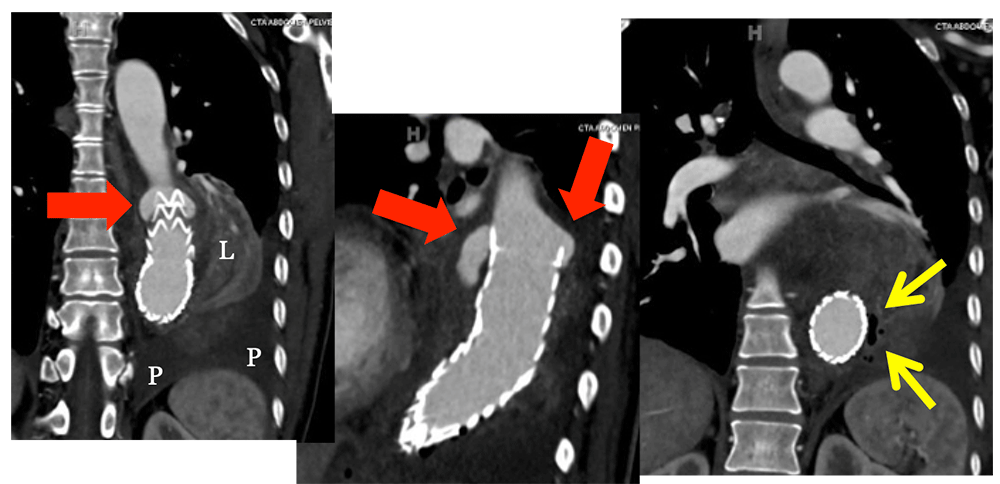
Aortoenteric Fistula

  • An endovascular stent does not protect against aneurysm sac leak or rupture if sac continues to expand in presence of endoleak.
  • In addition to frank rupture, the aneurysm sac may fistulize to adjacent bowel, commonly the duodenum as it drapes over the sac.
  • Often the patient experiences a herald bleed that brings them to the emergency department, which is intermittent so that active bleeding from the aneurysm sac is not seen at the time of imaging.
  • While contrast extravasation from the aorta to the bowel is not often NOT visualized in these cases, it provides definitive diagnosis when identified and mandates immediate intervention.
  • Adherence of the thickened colon to the anterior sac wall and stranding are clues to the diagnosis, strengthened by the presence of gas in the aneurysm sac.


Aortoduodenal Fistula

Patient with fractured stent (yellow arrow) status post replacement stent. Two years later, the patient presented to ED with melena due to aortoduodenal fistula from the aneurysm sac (red arrows).

Aortoduodenal Fistula


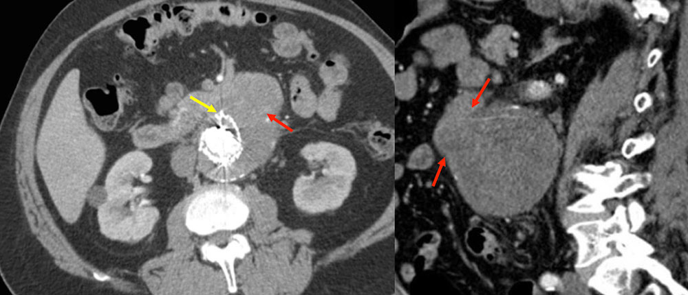
Aortoduodenal Fistula

87 year old female with CAD, AAA s/p endoluminal repair and revision who presented with progressive weakness. She was found to be severely anemic with baseline hemoglobin of 6 due to fistula between aneurysm sac and adjacent duodenum. In this case, gas bubbles (red arrows) are identified in the aneurysm sac, confirming fistulous connection (yellow arrows) to the adjacent duodenum. Heterogeneity within the sac reflects hematoma due to sac rupture.

Aortoduodenal Fistula


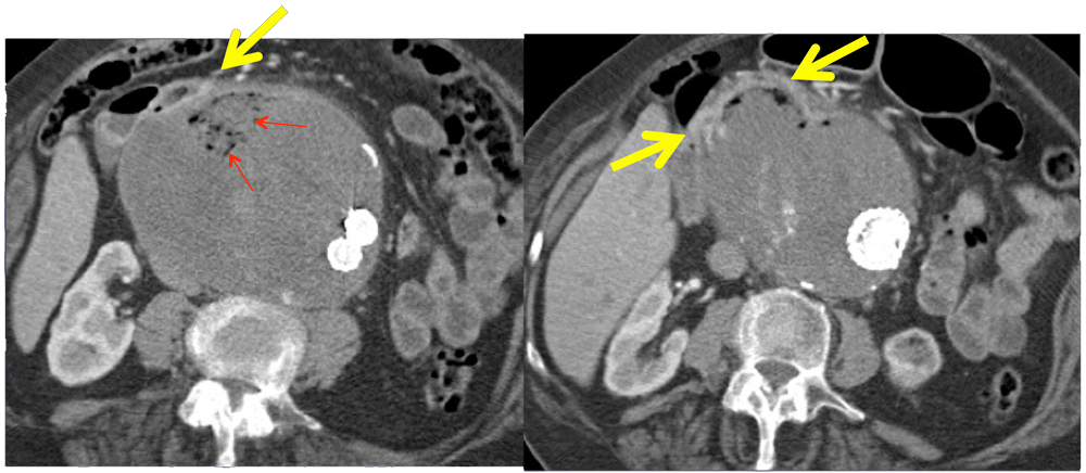
Aortocolonic Fistula

78 year old male s/p aortoiliac EVAR with chronic occlusion of left limb and chronic endoleak who presented with LGIB. CECT revealed aortocolonic fistula (arrows) from transverse colon to anterior sac.

Aortocolonic Fistula


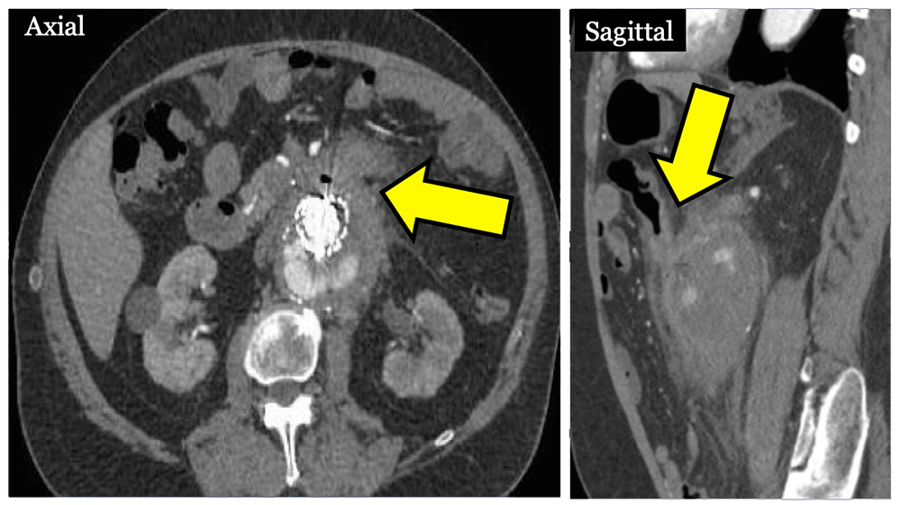
Retrograde Aortic Dissection

  • Rare catastrophic complications include retrograde aortic dissections
  • Sanford type A dissection (retrograde ascending aorta) after thoracic EVAR
    • Incidence ranges from 2-6%
    • Cause is often related to the procedure itself
    • Associated with the use of proximal bare spring stent grafts, previous type B dissection
    • Can be asymptomatic in up to 25%, underscoring importance of surveillance
  • Sanford Type B dissection after repair of AAA
    • Very rare, typically with fatal outcomes
  • Pre-existing atherosclerotic disease including ulcerated plaques or ectasia increase risk
  • Other etiologies likely related to difficulties in stent placement
    • Intimal calcifications
    • Angulated necks
    • Oversizing stents
    • Unstable plaques


TEVAR with Retrograde Dissection

82 year old female status post aortic arch and descending thoracic EVAR who presented with cardiac arrest due to ruptured retrograde Type A dissection (circle). Note extensive mediastinal hematoma (H) markedly narrowing the pulmonary arteries. This is a potential complication of a stent positioned in the aortic arch.
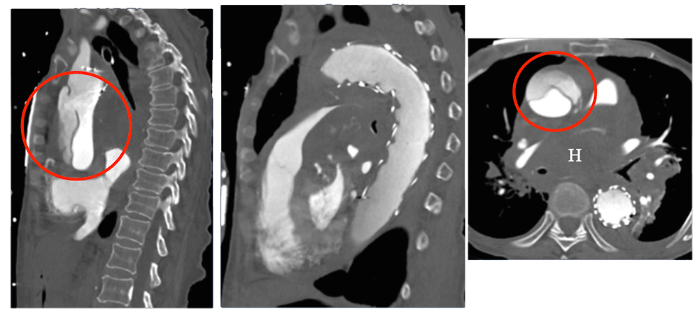
TEVAR with Retrograde Dissection


Rupture

  • Delayed rupture is rare, ranging from 0.5% to 2% per year.
  • Refractory or occult Type II endoleaks and Type V endoleaks increase sac pressure leading to rupture
  • May also occur without increasing aneurysm size
    • Likely due to sudden pressurization from type I/III endoleak
  • Imaging findings are important for treatment planning
    • Redo EVAR vs open repair


Ruptured Aneurysm Sac

Post AAA EVAR with endoleak seen best on venous phase (large arrow), which led to aneurysm sac rupture (small arrows).
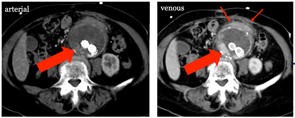
Ruptured Aneurysm Sac


References

  • Endovascular versus Open Repair of Abdominal Aortic Aneurysm. N Engl J Med 2010 05/20; 2016/08;362(20):1863-1871.
  • Arko III FR, Murphy EH, Boyes C, Nussbaum T, Lalka SG, Holleman J, et al. Current Status of Endovascular Aneurysm Repair: 20 Years of Learning. Semin Vasc Surg 2012 9;25(3):131-135.
  • Becquemin J, Allaire E, Desgranges P, Kobeiter H. Delayed Complications Following EVAR. Techniques in Vascular and Interventional Radiology 2005 3;8(1):30-40.
  • Blankensteijn JD, de Jong SECA, Prinssen M, van dH, Buth J, van Sterkenburg SMM, et al. Two-Year Outcomes after Conventional or Endovascular Repair of Abdominal Aortic Aneurysms. N Engl J Med 2005 06/09; 2016/08;352(23):2398-2405.
  • Brown A, Saggu GK, Bown MJ, Sayers RD, Sidloff DA. Type II endoleaks: challenges and solutions. Vascular Health and Risk Management 2016 03/02;12:53-63.
  • Bryce Y, Rogoff P, Romanelli D, Reichle R. Endovascular Repair of Abdominal Aortic Aneurysms: Vascular Anatomy, Device Selection, Procedure, and Procedure-specific Complications. Radiographics 2015 03/01; 2016/08;35(2):593-615.


References

  • Catanescu I, Long G, Bove P, Khoury M, Brown O, Rimar S, et al. Rupture of Abdominal Aortic Aneurysm in Patients with and without Antecedent Endovascular Repair. Ann Vasc Surg .
  • Conrad MF, Adams AB, Guest JM, Paruchuri VB,David C., LaMuraglia GM, Cambria RP. Secondary Intervention After Endovascular Abdominal Aortic Aneurysm Repair. Ann Surg 2009 September;250(3):383-389.
  • De Bruin JL, Baas AF, Buth J, Prinssen M, Verhoeven ELG, Cuypers PWM, et al. Long-Term Outcome of Open or Endovascular Repair of Abdominal Aortic Aneurysm. N Engl J Med 2010 05/20; 2016/08;362(20):1881-1889.
  • Eggebrecht H, Thompson M, Rousseau H, Czerny M, Lönn L, Mehta RH, et al. Retrograde Ascending Aortic Dissection During or After Thoracic Aortic Stent Graft Placement. Circulation 2009 09/14;120(11):S276.
  • Khanbhai M, Ghosh J, Ashleigh R, Baguneid M. Type B aortic dissection after standard endovascular repair of abdominal aortic aneurysm. BMJ Case Reports 2013 May 09;2013.


References

  • Lal BK, Zhou W, Li Z, Kyriakides T, Matsumura J, Lederle FA, et al. Predictors and outcomes of endoleaks in the Veterans Affairs Open Versus Endovascular Repair (OVER) Trial of Abdominal Aortic Aneurysms. Journal of Vascular Surgery 2015 12;62(6):1394-1404.
  • Maldonado TS, Rockman CB, Riles E, Douglas D, Adelman MA, Jacobowitz GR, et al. Ischemic complications after endovascular abdominal aortic aneurysm repair. Journal of Vascular Surgery 2004 10;40(4):703-710.
  • Mehta M, Sternbach Y, Taggert JB, Kreienberg PB, Roddy SP, Paty PSK, et al. Long-term outcomes of secondary procedures after endovascular aneurysm repair. Journal of Vascular Surgery 2010 12;52(6):1442-1449.
  • Mertens J, Houthoofd S, Daenens K, Fourneau I, Maleux G, Lerut P, et al. Long-term results after endovascular abdominal aortic aneurysm repair using the Cook Zenith endograft. Journal of Vascular Surgery 2011 7;54(1):48-57.e2.
  • Murphy EH, Szeto WY, Herdrich BJ, Jackson BM, Wang GJ, Bavaria JE, et al. The management of endograft infections following endovascular thoracic and abdominal aneurysm repair. Journal of Vascular Surgery 2013 11;58(5):1179-1185.


References

  • Schermerhorn ML, Buck DB, O’Malley AJ, Curran T, McCallum JC, Darling J, et al. Long-Term Outcomes of Abdominal Aortic Aneurysm in the Medicare Population. N Engl J Med 2015 07/23; 2016/08;373(4):328-338.
  • Sirignano P, Pranteda C, Capoccia L, Menna D, Mansour W, Speziale F. Retrograde Type B Aortic Dissection as a Complication of Standard Endovascular Aortic Repair. Ann Vasc Surg 2015 1;29(1):127.e5-127.e9.
  • Stavropoulos SW, Charagundla SR. Imaging Techniques for Detection and Management of Endoleaks after Endovascular Aortic Aneurysm Repair. Radiology 2007 06/01; 2016/08;243(3):641-655.
  • Vandy F, Upchurch GR. Endovascular Aneurysm Repair: Current Status. Circulation: Cardiovascular Interventions 2012 December 01;5(6):871-882.


References

  • Wadgaonkar AD, Black JH, Weihe EK, Zimmerman SL, Fishman EK, Johnson PT. Abdominal Aortic Aneurysms Revisited: MDCT with Multiplanar Reconstructions for Identifying Indicators of Instability in the Pre- and Postoperative Patient. Radiographics 2015 01/01; 2016/08;35(1):254-268.
  • Walker TG, Kalva SP, Yeddula K, Wicky S, Kundu S, Drescher P, et al. Clinical Practice Guidelines for Endovascular Abdominal Aortic Aneurysm Repair: Written by the Standards of Practice Committee for the Society of Interventional Radiology and Endorsed by the Cardiovascular and Interventional Radiological Society of Europe and the Canadian Interventional Radiology Association. Journal of Vascular and Interventional Radiology 2010 11;21(11):1632-1655.

Privacy Policy

Copyright © 2024 The Johns Hopkins University, The Johns Hopkins Hospital, and The Johns Hopkins Health System Corporation. All rights reserved.