Potential Clinical Applications of the Cinematic Rendering Virtual Topogram
Potential Clinical Applications of the Cinematic Rendering Virtual Topogram Linda C. Chu The Russel H. Morgan Department of Radiology and Radiological Science, Johns Hopkins University, Baltimore |
Disclosure:
|
Overview
|
Cinematic Rendering
|
Volume Rendering (VR) vs. Cinematic Rendering (CR) 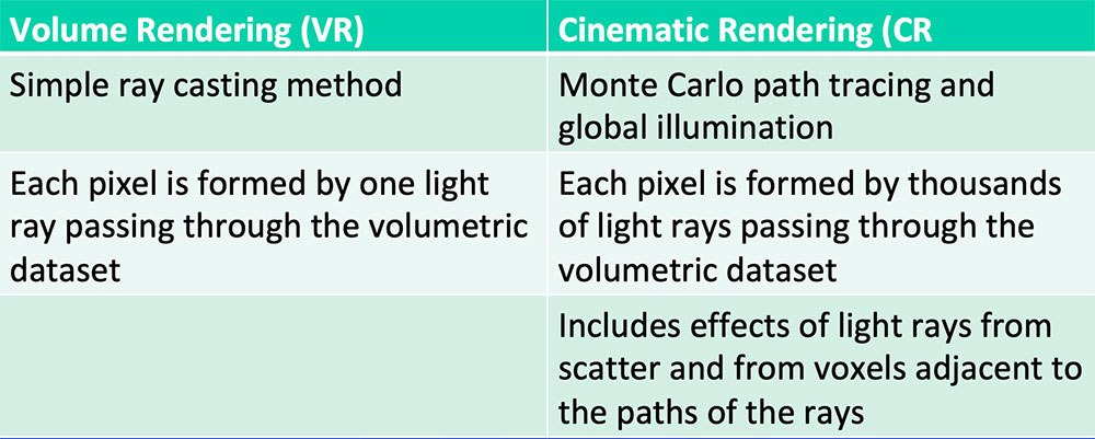 Comaniciu D et al. Med Image Anal. 2016;33:19-26. Fellner FA et al. J Biomed Sci Eng. 2016;9:170-5. Eid M et al. AJR. 2017;209(2):370-9. Johnson PT et al. AJR. 2017;209(2):309-12. |
Traditional Topogram
 Traditional Topogram Johnson PT et al. AJR. 2014;202(6):1256-1263. |
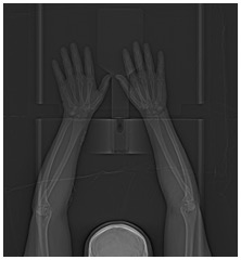
Virtual Topogram
|
Virtual Topogram Virtual topogram with skin, soft tissue, and bone presets 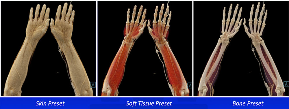 Chu LC et al. Emerg Radiol. 2019;26(5):573-580 |
Potential Applications
|
Skin and Soft Tissue Pathology
|
Trauma 36-year-old man with gunshot wound to the left thigh  A) Coronal IV contrast-enhanced CT of the left thigh with CR soft tissue preset shows a focal soft tissue defect from gunshot wound. B) CR in vascular and soft tissue preset shows the underlying vascular injury with transection and occlusion of the left superficial femoral artery. C) CR in vascular preset fades out the soft tissues and highlights the vascular injury. |
Trauma 52-year-old man involved in a motor vehicle accident 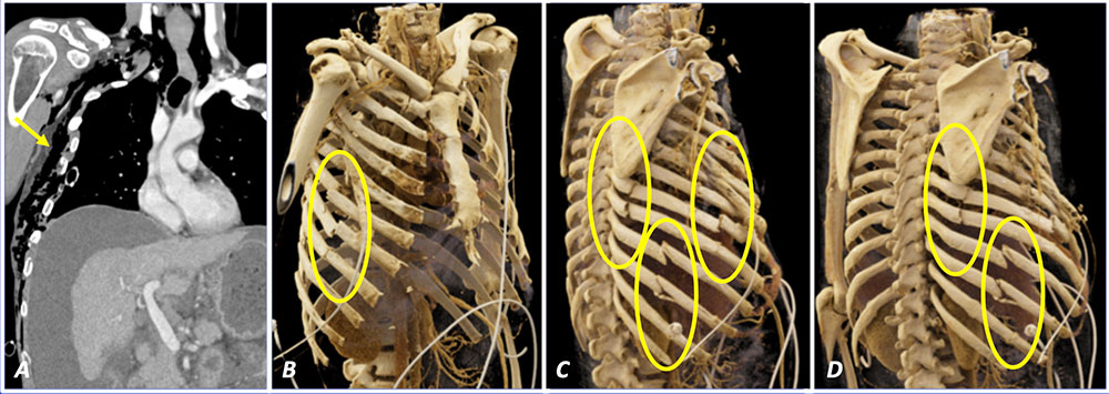 A) Coronal IV contrast-enhanced CT of the chest shows extensive right chest wall deformity and subcutaneous emphysema. B-D) CR in bone presets in varying obliquities better illustrates the extent of numerous right rib fractures than the 2D image. They allow for comprehensive and efficient assessment of osseous injuries in a trauma patient. |
Soft Tissue Infection 31-year-old woman with right arm infection and clinical concern for abscess 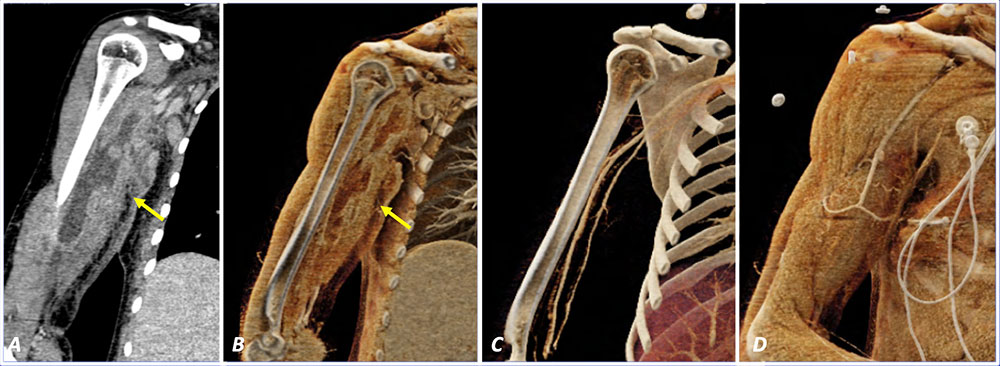 A) Coronal IV contrast-enhanced CT of the right arm shows a loculated peripherally enhancing fluid collection in the medial right arm consistent with abscess. B) Coronal CR in soft tissue preset similarly shows the extent the abscess. C) CR in vascular preset shows patency of right arm arteries with no evidence of compromise. D) CR soft tissue topogram can be used for anatomic localization of the fluid collection for percutaneous drainage. |
Soft Tissue Infection 36-year-old man with recent left thigh laceration repair who presents with increased thigh swelling 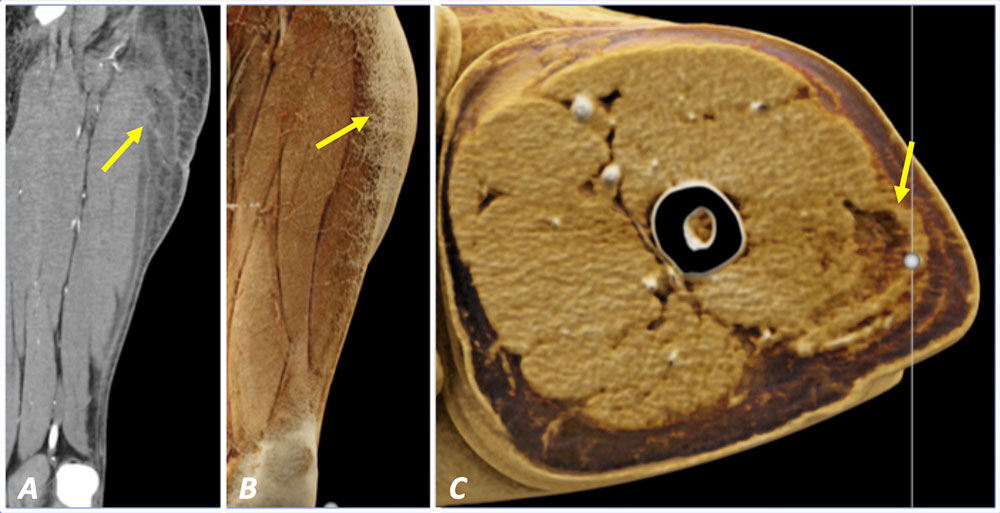 A) Coronal IV contrast-enhanced CT of the left thigh shows a loculated peripherally enhancing fluid collection in the lateral left thigh consistent with abscess. B) Coronal CR in soft tissue preset accentuates diffuse subcutaneous edema in the left thigh and can help localize the region of interest. C) Axial CR in soft tissue preset highlights difference in texture and attenuation of the abscess relative to background soft tissues. |
Soft Tissue Injection Granulomas 33 year-old woman with abdomen pain 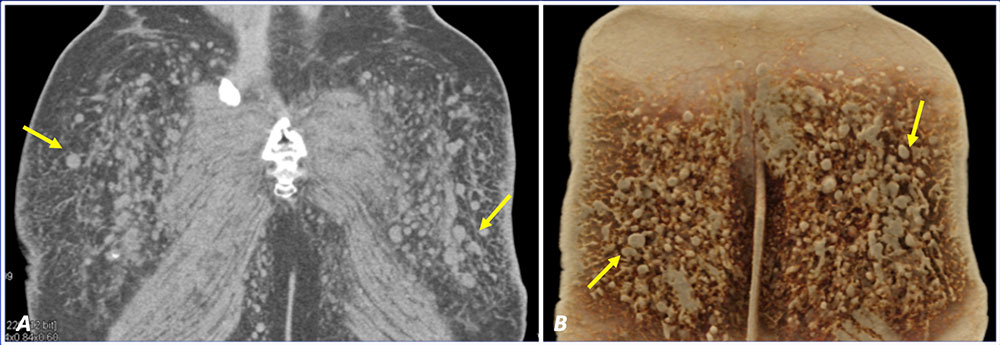 A) Coronal IV contrast-enhanced CT of the pelvis shows an innumerable granulomas within bilateral gluteal soft tissues, presumably related to cosmetic procedures. B) Coronal CR in soft tissue preset accentuates the textural difference between the granulomas and subcutaneous fat. |
Soft Tissue Metastasis 74-year-old man with metastatic melanoma to the right axilla 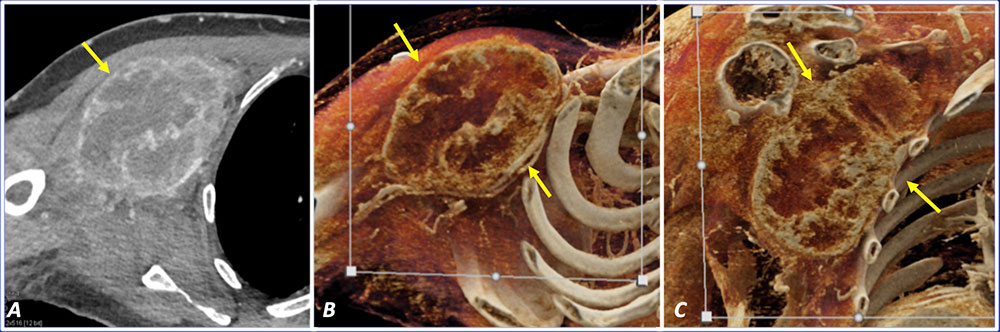 A) Axial IV contrast-enhanced CT of the chest shows heterogeneous enhancing mass in the right axilla. B) Axial CR in soft tissue preset highlights the textural difference of the right axillary mass and compression of the right axillary vasculature. C) Coronal CR in soft tissue preset shows the close proximity of the right axillary mass to the underlying chest wall. |
Soft Tissue Metastasis 57-year-old man with history of metastatic lung cancer 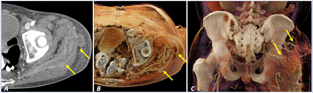 A) Axial IV contrast-enhanced CT of the pelvis shows an infiltrative enhancing mass within the left gluteus musculature and subcutaneous soft tissues. B) Axial CR in soft tissue preset improves visualization of the soft tissue mass by accentuating textural difference. C) Coronal CR in vascular preset shows prominent neovascularity supplying the soft tissue metastasis. |
Abdominal Wall Defect 50-year-old woman with Crohns disease status post bowel resection with open abdominal wall wound 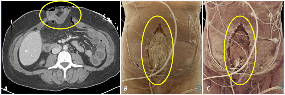 A) Axial IV contrast-enhanced CT of the abdomen shows an open abdomen wall wound with small bowel herniation. B) Coronal CR in skin preset shows the extent of the abdominal wall defect and the overlying wound vacuum device. C) Coronal CR in soft tissue preset shows the underlying small bowel by altering the transparency of the CR settings. |
Abdominal Wall Ostomy 35-year-old paraplegic man with neurogenic bowel with left lower quadrant colostomy and chronic DVTs 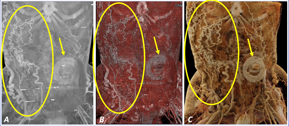 A) Coronal IV contrast-enhanced CT in maximum intensity projection (MIP) shows the left lower quadrant ostomy and prominent superficial venous collaterals from chronic DVT. B) Coronal VR illustrates the same findings with slight improvement in soft tissue differentiation. C) Coronal CR in soft tissue and vascular preset improve depth perception and visualization of these findings. |
Superficial Clues to Deeper Pathology
|
Differential Diagnosis? Neuroendocrine tumors Gastrointestinal stromal tumors Hypervascular metastases 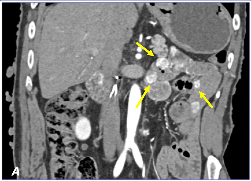 A) Coronal IV contrast-enhanced CT of the abdomen shows multiple enhancing jejunal masses. |
Neurofibromatosis 1 57 year-old man with neurofibromatosis 1 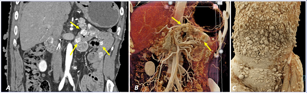 A) Coronal IV contrast-enhanced CT of the abdomen shows multiple enhancing jejunal masses. B) Coronal CR similarly shows multiple enhancing jejunal masses. C) Coronal CR in skin preset highlights innumerable neurofibromas in the skin, consistent with neurofibromatosis 1. This clue helps to suggest the correct diagnosis of multiple gastrointestinal stromal tumors (GISTs) in the setting of neurofibromatosis 1. |
Neurofibromatosis 1 33 year-old man with neurofibromatosis 1 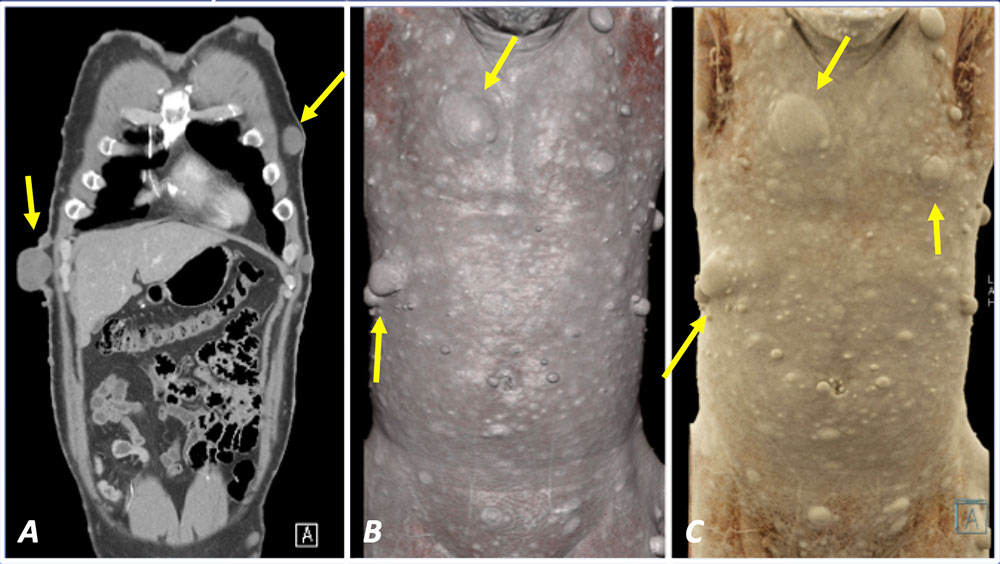 A) Coronal IV contrast-enhanced CT shows numerous subcutaneous nodules consistent with neurofibromas in the setting of neurofibromatosis 1. B) Coronal VR similarly shows multiple neurofibromas. C) Coronal CR in skin preset accentuates subtle irregularities of the skin surface compared to traditional volume rendering and can improve visualization of skin pathology compared to 2D and volume rendering. |
Aortic Occlusion 56 year-old man with chronic aortic occlusion and bilateral axillary-femoral bypass grafts 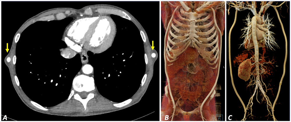 A) Axial IV contrast-enhanced CT partially demonstrate patent bilateral axillary-femoral grafts. B) Coronal CR in vascular soft tissue preset shows patency of bilateral axillary-femoral grafts. C) Coronal CR in vascular preset viewed in the posterior projection clearly define patency of bilateral axillary-femoral grafts and evidence of old occluded aortoiliac stents. |
Aortic Occlusion 69 year-old woman with chronic aortic occlusive disease with occluded axillary-femoral and femoral-femoral bypass grafts 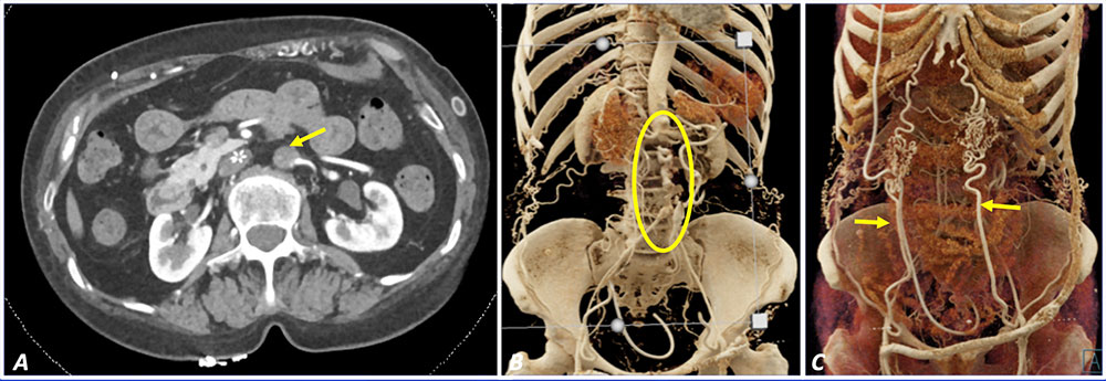 A) Axial IV contrast-enhanced CT shows chronic occlusion of infrarenal abdominal aorta. B) Coronal CR in vascular preset shows chronic occlusion of infrarenal aorta with reconstitution of the iliac arteries via epigastric collaterals. C) Coronal CR in vascular preset focused on superficial tissues highlight the hypertrophy of the epigastric arteries as important sources of collateralization. These collaterals are better appreciated on CR compared to 2D images. |
SVC Occlusion 39 year-old man with chronic SVC occlusion 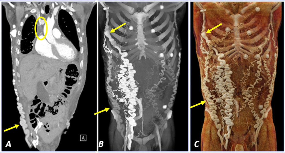 A) Coronal IV contrast-enhanced CT shows chronic occlusion of SVC with prominent body wall collaterals. B) Coronal MIP better illustrates the extent of body wall collaterals compared to 2D image. C) Coronal CR in vascular and soft tissue preset shows superior soft tissue differentiation compared to MIP and portrays the findings with increased detail. |
Portal Hypertension 54 year-old man with cirrhosis and portal hypertension 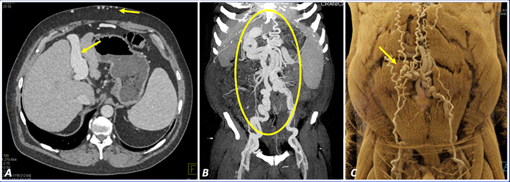 A) Axial IV contrast-enhanced CT cirrhotic liver with recanalization of paraumbilical vein and engorgement of superficial venous collaterals, consistent with portal hypertension. B) Coronal MIP better illustrates the extent of venous collaterals compared to 2D image. C) Coronal CR in vascular and soft tissue preset improve depth perception and highlight the engorged superficial epigastric veins, with classic appearance of caput medusae. |
Treatment Planning
|
Fracture Blisters 60-year-old man with right humerus fracture complicated by fracture blisters 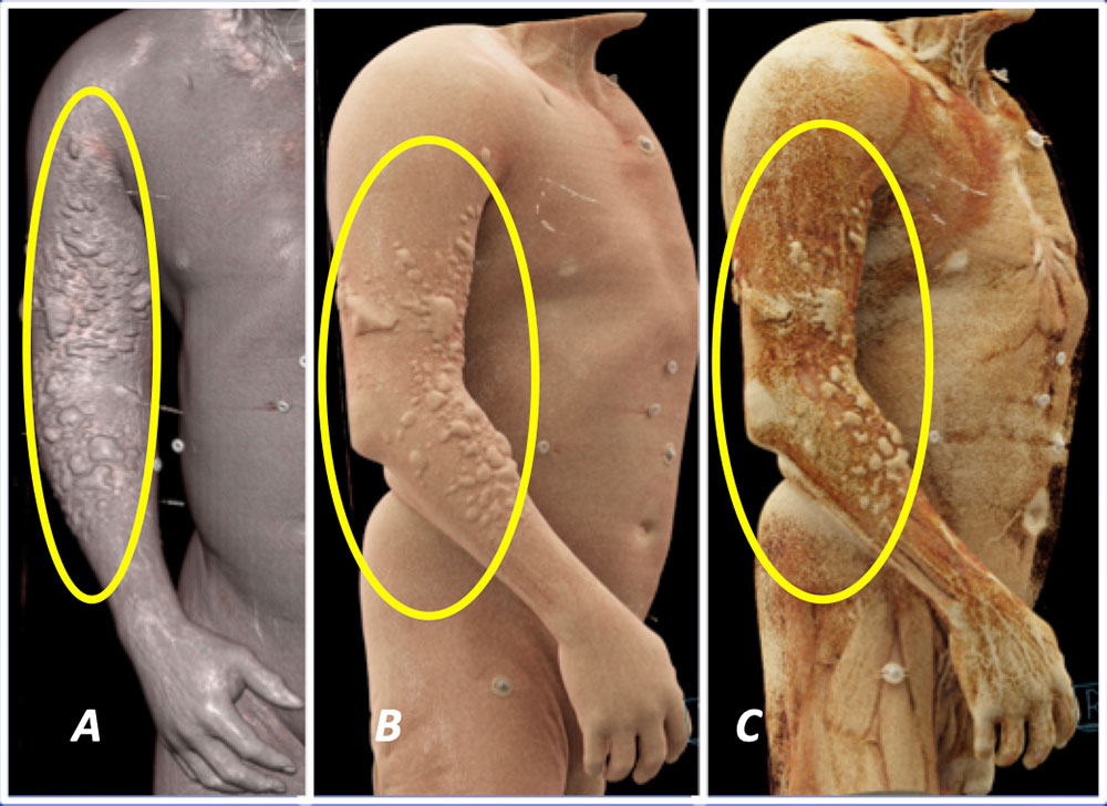 A) Coronal VR IV contrast-enhanced CT of the right arm shows multiple raised areas in the skin compatible with fracture blisters. B) Sagittal oblique CR in skin preset and C) Sagittal oblique CR in soft tissue preset improve the visualization of the skin blisters due to improved soft tissue differentiation and depth perception. |
Repair of Traumatic Injuries 32-year-old man with traumatic right calf laceration 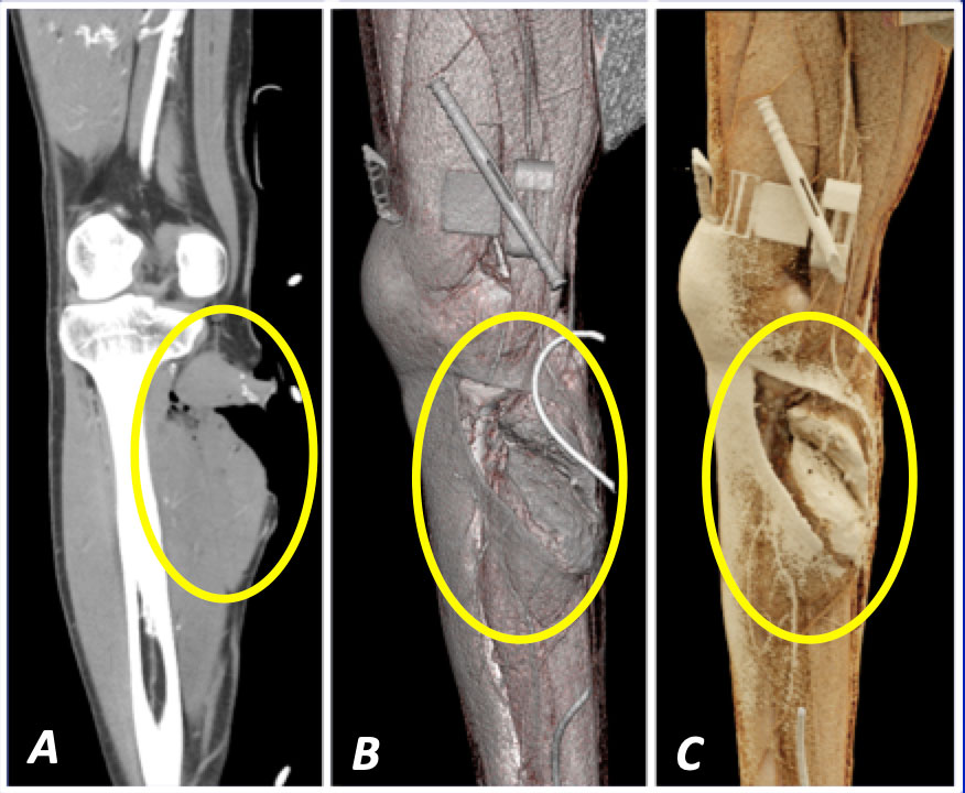 A) Coronal IV contrast-enhanced CT of the right calf shows a deep laceration along the medial right calf. B) Sagittal oblique VR again demonstrates the deep laceration. C) Sagittal oblique CR in soft tissue preset improves soft tissue differentiation and visualization of the underlying fascial and muscle laceration. |
Repair of Traumatic Injuries 27-year-old man with traumatic right wrist laceration 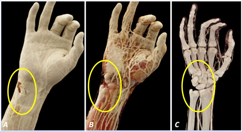 A) Coronal IV contrast-enhanced CT of the right wrist in CR skin preset shows a laceration along ulnar aspect of the wrist. B) CR in soft tissue preset shows integrity of the underlying tendons without evidence of acute injury. C) CR in vascular preset shows absence of vascular injury. |
Venous Malformation 39-year-old woman with left gluteal venous malformation who presents for pre-operative planning 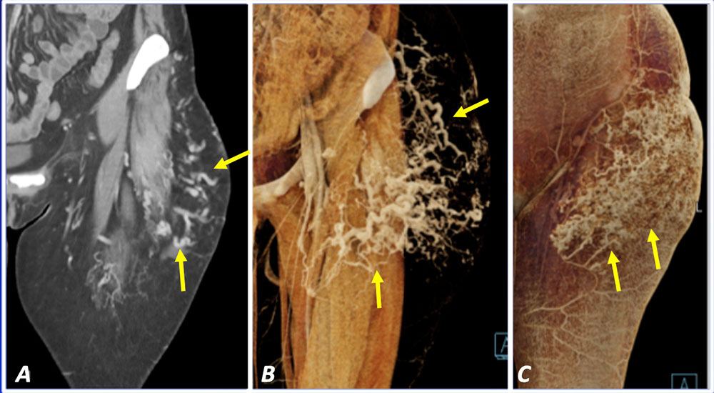 A) Coronal IV contrast-enhanced CT of the left thigh shows a few dilated veins. Full extent of the venous malformation is difficult to appreciate on 2D images. B) Coronal CR in soft tissue and vascular preset shows the extensive venous malformation and relationship with the underlying soft tissues. C) Coronal CR in soft tissue preset highlights the superficial cutaneous findings, which can be used for localization of the deeper venous malformation. |
Limitations
|
Conclusion
|
References
Acknowledgements
|