Cinematic Rendering of Liver Diseases – Preliminary Observations
Cinematic Rendering of Liver Diseases – Preliminary Observations Linda C. Chu The Russell H. Morgan Department of Radiology and Radiological Science, Johns Hopkins University, Baltimore, MD |
Disclosures
|
Learning Objectives
|
Cinematic Rendering (CR)
 Comaniciu D et al. Med Image Anal. 2016;33:19-26. Fellner FA et al. J Biomed Sci Eng. 2016;9:170-5. Eid M et al. AJR. 2017;209(2):370-9. Johnson PT et al. AJR. 2017;209(2):309-12. |
Volume Rendering (VR) vs. Cinematic Rendering (CR)
 Calhoun PS et al. Radiographics. 1999;19(3):745-64. Fishman EK et al. Radiographics. 2006;26(3):905-22. |
Volume Rendering (VR) vs. Cinematic Rendering (CR) Volume Rendering (VR)
Eid M et al. AJR. 2017;209(2):370-9. Johnson PT et al. AJR. 2017;209(2):309-12. |
Cinematic Rendering (CR)
 Rowe SP et al. JCCT. 2018;12(1):56-59. Rowe SP et al. Emerg Radiol. 2018;25(1):93-101. Chu LC et al. Abdom Radiol (NY). 2018 [Epub ahead of print]. Johnson PT et al. AJR. 2017;209(2):309-312. |
Case Review of CR in Liver Imaging Focal Liver Masses
|
Case 1: Hepatic Hemangioma 49 year-old woman with hepatic hemangioma. 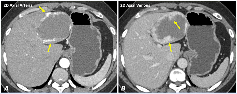 Axial IV contrast enhanced CT images show peripheral nodular enhancement (A) with progressive centripetal filling (B) of a classic hepatic hemangioma. |
Case 1: Hepatic Hemangioma 49 year-old woman with hepatic hemangioma. 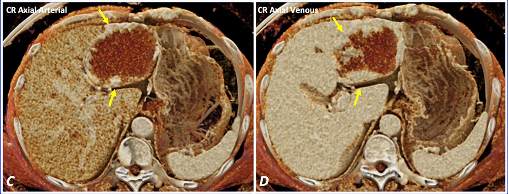 Cinematic rendering (C and D) can accentuate the peripheral nodular enhancement pattern compared to 2D axial images (A and B). |
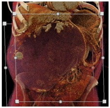
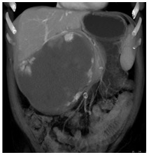
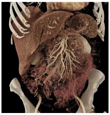
Case 2: Giant Hepatic Hemangioma 42 year-old woman with a giant hepatic hemangioma. 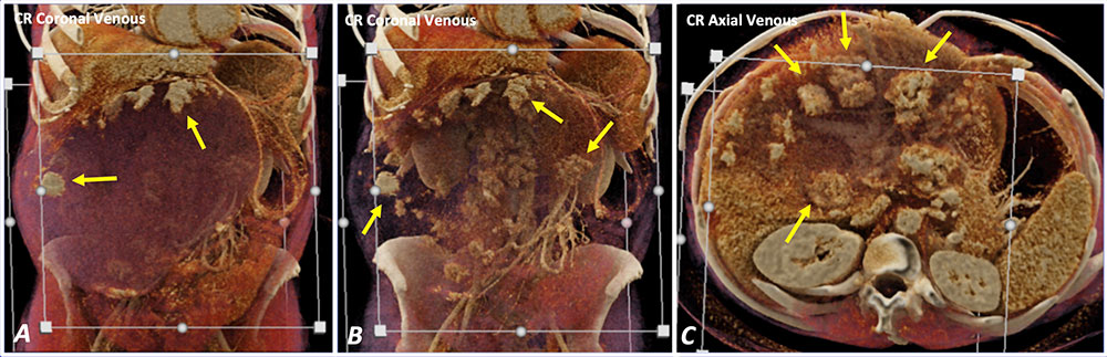 Cinematic rendering shows classic peripheral nodular enhancement of a giant hepatic hemangioma (A). Translucency of the mass can be altered with the dynamic display (B and C) to appreciate this enhancement pattern. |
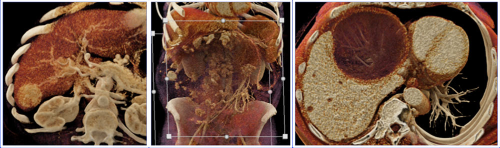
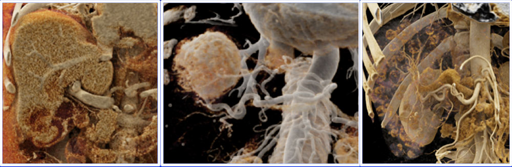
Case 3: Focal Nodular Hyperplasia 52 year-old woman with no prior liver disease with incidentally diagnosed focal nodular hyperplasia. 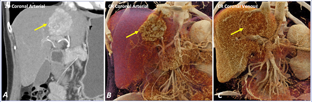 Arterial phase coronal IV contrast enhanced CT image (A) shows a homogeneously hypervascular mass in the left hepatic lobe. Arterial phase CR image (B) shows increased conspicuity of the mass compared to 2D images. Venous phase CR image (C) shows isoenhancement of the mass relative to background liver. |
Case 3: Focal Nodular Hyperplasia 52 year-old woman with no prior liver disease with incidentally diagnosed focal nodular hyperplasia. 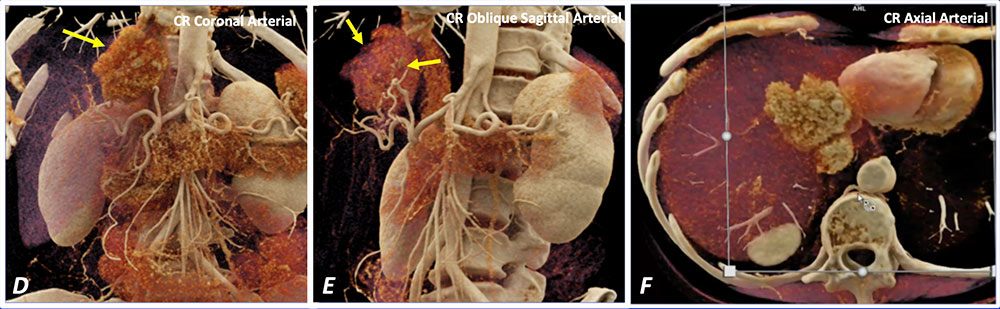 Background liver can be rendered translucent to increase conspicuity of the hypervascular mass (D). CR vascular map (E) can highlight the central feeding artery and spokewheel branches, classic features of focal nodular hyperplasia. CR video (F) shows manipulation of the dynamic window display. |
Case 4: Hepatic Adenoma 32 year-old woman with no prior liver disease with a hypervascular liver mass. Resection showed hepatic adenoma. 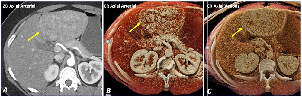 Axial IV contrast enhanced CT image (A) shows a heterogeneously enhancing left hepatic lobe mass in a non-cirrhotic liver. CR images can accentuate the enhancement on both arterial and venous phases (B and C) to increase tumor conspicuity. |
Case 5: Liver Abscess 70 year-old man with sepsis and liver abscess. Fine needle aspiration returned scant fluid positive for Gram negative bacteria. 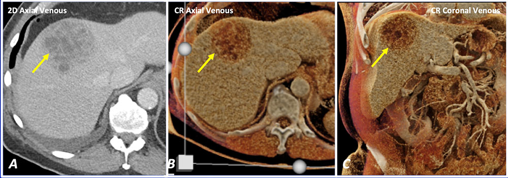 Axial IV contrast enhanced CT image (A) shows a multiseptated cystic mass in the liver dome. CR images (B and C) can increase the appreciation of the thick enhancing internal septations, a feature that suggests the mass may be difficult to drain with percutaneous approach. |
Case 6: Liver Abscess 49 year-old woman with liver abscess s/p pigtail placement. 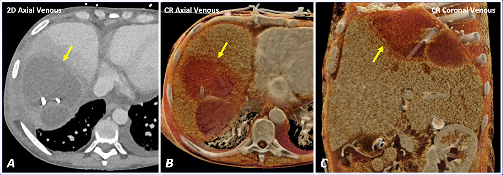 Axial IV contrast enhanced CT image (A) shows a peripherally enhancing cystic mass within the liver dome. CR images (B and C) can increase the appreciation of the thick enhancing capsule. |
Case 7: Hepatocellular Carcinoma 79 year-old man with history of hepatitis B and C with hepatocellular carcinoma. 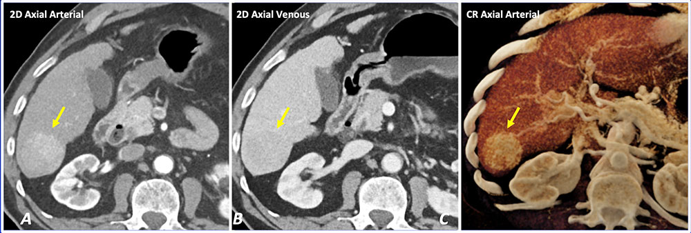 Axial IV contrast enhanced CT images (A and B) show a subtle hypervascular mass (A) with subtle washout on venous phase (B). CR (C) can increase the conspicuity of the subtle hypervascular mass. |
Case 8: Hepatocellular Carcinoma 84 year-old woman with biopsy proven hepatocellular carcinoma. 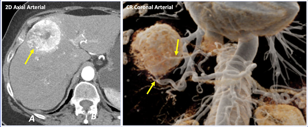 Axial IV contrast enhanced CT image (A) shows a heterogeneous hypervascular mass in the right hepatic lobe. CR images (B) can render the background liver transparent to highlight the feeding arteries, and can potentially help in pretreatment planning for chemoembolization. |
Case 9: Cholangiocarcinoma 75 year-old woman with biopsy proven cholangiocarcinoma. 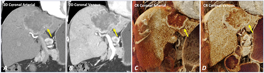 Coronal IV contrast enhanced CT images (A and B) show a heterogeneously enhancing liver mass (A) with progressive delayed enhancement (B). CR images (C and D) accentuate the heterogeneous internal enhancement – an important feature in distinguishing malignant from benign liver masses. |
Case 10: Cholangiocarcinoma 60 year-old woman with history of cholangiocarcinoma. 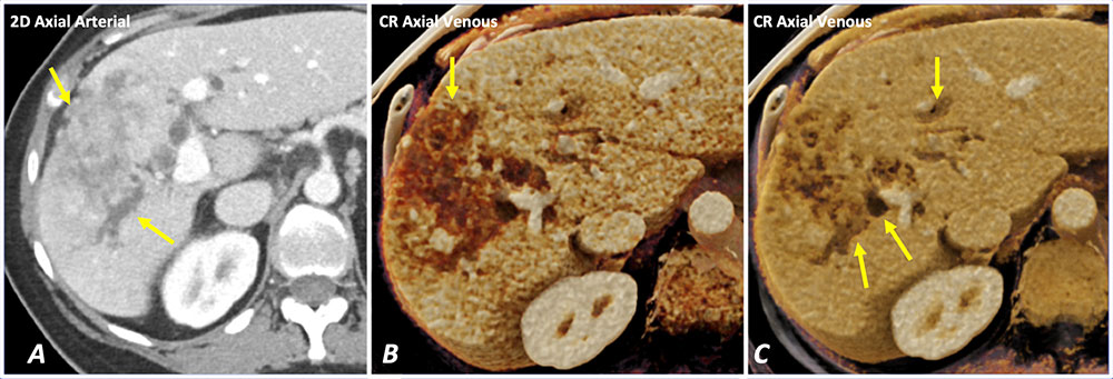 Axial IV contrast enhanced CT image (A) shows a heterogeneously enhancing mass with intrahepatic biliary ductal dilatation. Dynamic window display of CR images (B and C) can be adjusted to highlight the mass (B) and the dilated bile ducts (C). |
Case 11: Mixed Cholangiocarcinoma and Hepatocellular Carcinoma 92 year-old man with biopsy-proven mixed cholangiocarcinoma and hepatocellular carcinoma with portal vein thrombosis. 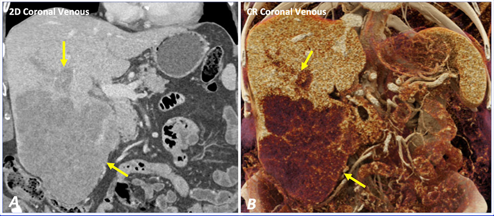 Coronal IV contrast enhanced CT image (A) shows a large hypoenhancing mass in the right hepatic lobe with thrombosis of portal vein branches. CR (B) can increase the conspicuity of the liver mass and portal vein thrombus. |
Case 12: Metastatic Cholangiocarcinoma 61 year-old woman with metastatic cholangiocarcinoma. 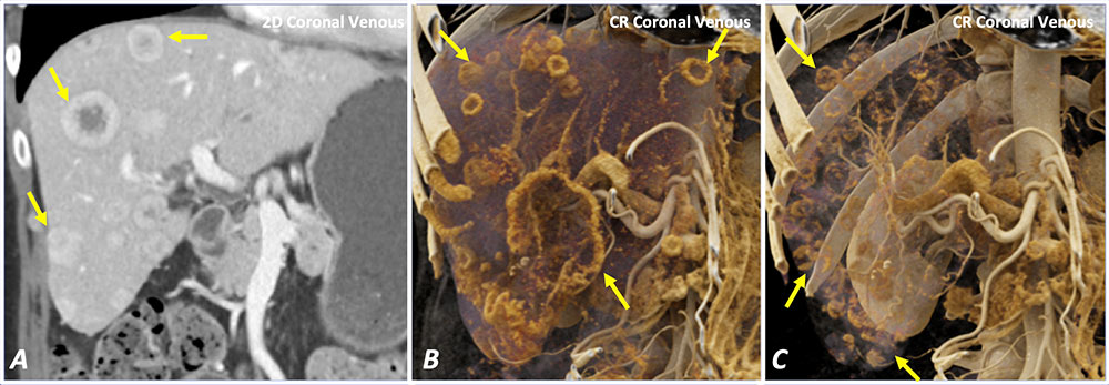 Coronal IV contrast enhanced CT image (A) shows numerous enhancing liver masses with targetoid appearance. CR images (B and C) can render the background liver translucent to highlight the global disease burden. |
Case 13: Metastatic Colon Cancer with Gallbladder Invasion 79 year-old man with history of metastatic colon cancer. 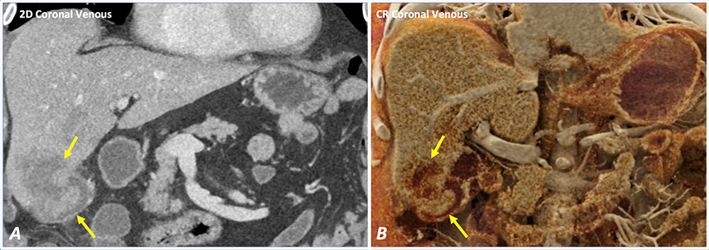 Coronal IV contrast enhanced CT image (A) shows a liver metastasis in the right hepatic lobe with invasion of the gallbladder lumen. CR (B) can improve depth perception and increase appreciation of tumor invasion into the gallbladder. |
Case 14: Metastatic GIST 58 year-old man with history of metastatic gastrointestinal stromal tumor. 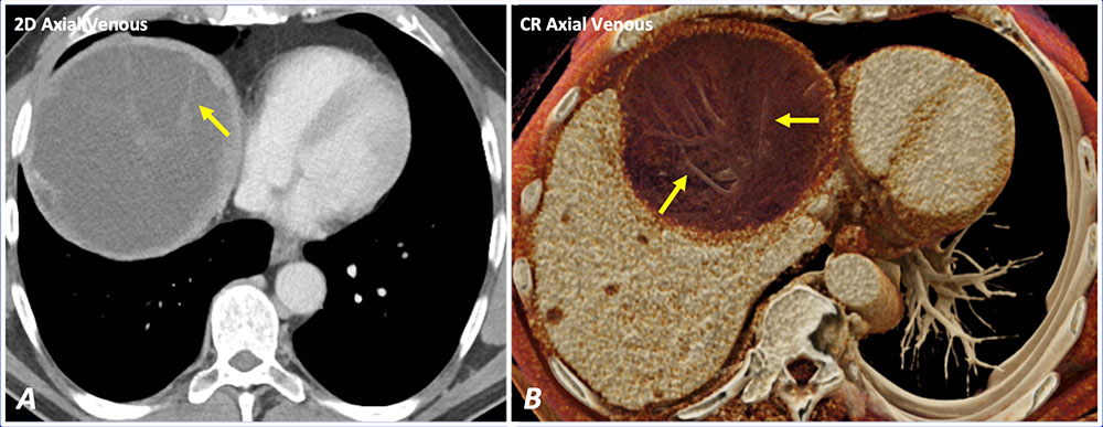 Axial IV contrast enhanced CT image (A) shows a cystic mass with multiple subtle internal septations. CR (B) improves the depth perception and appreciation of subtle internal septations and internal architecture. |
Case 15: Metastatic Head and Neck Squamous Cell Carcinoma 45 year-old man with metastatic head and neck squamous cell carcinoma. 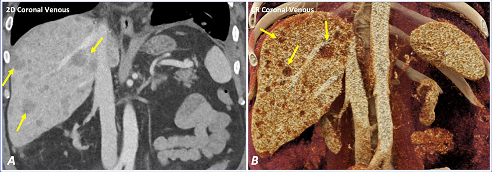 Coronal IV contrast enhanced CT image (A) shows numerous hypoenhancing liver lesions compatible with metastatic disease. CR image (B) can increase tissue contrast and increase the conspicuity of these metastatic lesions. |
Potential Benefits of CR in Evaluation of Focal Liver Masses
 |
Potential Benefits of CR in Evaluation of Focal Liver Masses
 |
Case 16: Cirrhosis 61 year-old man with history of cirrhosis. 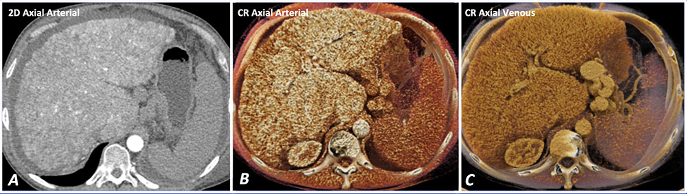 Axial IV contrast enhanced CT image (A) shows diffuse nodular parenchymal appearance of a cirrhotic liver. CR images (B and C) show greater appreciation of the nodular parenchymal texture due to improved depth perception and photorealism with CR. |
Case 17: Cirrhosis with Portal Hypertension 64 year-old woman with history of cirrhosis and portal hypertension.  CR allows the user to alter the window settings to highlight the anatomy of interest. In this case, the extensive portal systemic collaterals within the left lower quadrant can be highlighted in the vascular map (B) by rendering the overlapping organs transparent. |
Case 18: Cirrhosis with Portal Hypertension 69 year-old woman with history of cirrhosis s/p liver transplant with mild intrahepatic biliary dilatation, splenomegaly, and varices. 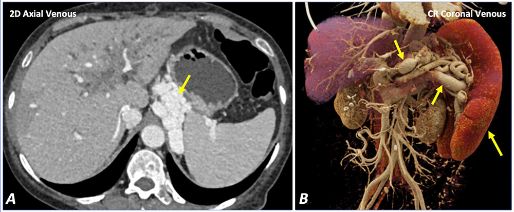 Axial IV contrast enhanced CT image (A) shows marked left upper quadrant varices from portal hypertension. CR image (B) can show the varices and splenomegaly with increased photorealistic detail. |
Discussion
|
Future Directions
|
Limitations
|
Conclusion
|
References
Acknowledgements
|