google ads

State-of-the-art Diagnosis and Treatment of Metastatic Melanoma: MDCT Protocol Optimization to Identify Metastases in Unusual Locations and Review of Innovative Therapeutic Agents

State-of-the-art Diagnosis and Treatment of Metastatic Melanoma: MDCT Protocol Optimization to Identify Metastases in Unusual Locations and Review of Innovative Therapeutic Agents

Elliot K. Fishman, M.D.

The Russell H. Morgan Department of Radiology and Radiological Science
The Johns Hopkins Medical Institutions
Baltimore, Maryland


Cardiac Metastasis

62-year-old man with melanoma metastatic to the right ventricle. Axial IV contrast enhanced CT with soft tissue (A) and high contrast (B) windows. Tumor conspicuity is enhanced by use of high contrast window (B).

Cardiac Metastasis


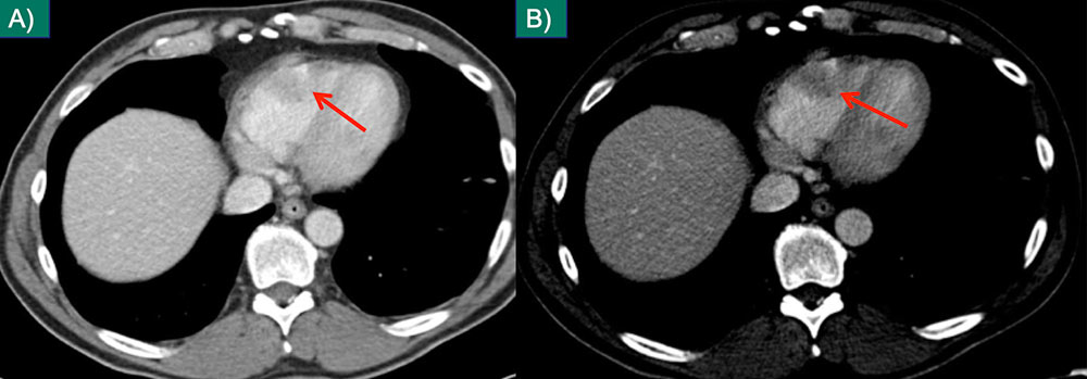
Intramuscular Metastases

55-year-old female with melanoma. Intramuscular metastasis (A) better visualized with high contrast window (B) in axial plane. Second muscular metastasis in the same patient on standard axial soft tissue window (C) and coronal MPR with contrast window (D).

Intramuscular Metastases


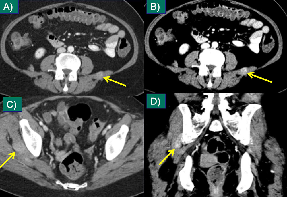
Spinal Canal

76-year-old male with melanoma; soft tissue infiltration in the sacral spinal canal (A) is better appreciated on the sagittal MPR with a soft tissue window (B), which also reveals cortical erosion of the posterior sacrum (C).

Spinal Canal


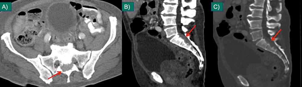
Parotid Metastasis

48-year old woman with melanoma and enhancing mass in the left parotid on axial (A) and coronal (B) IV contrast-enhanced CT. Metastases to solid organs are best visualized with high contrast windows (B). Confirmation with coronal MPR increases diagnostic confidence.

Parotid Metastasis


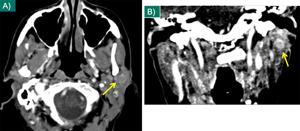
Thyroid Metastasis

55-year-old female with melanoma and thyroid metastasis seen on axial (A) and coronal (B) IV contrast-enhanced CT.

Thyroid Metastasis


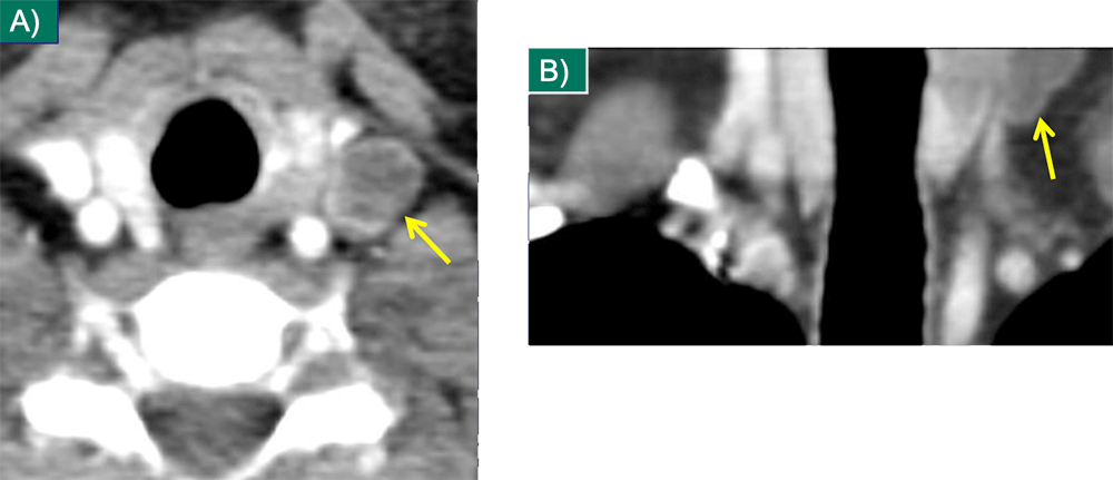
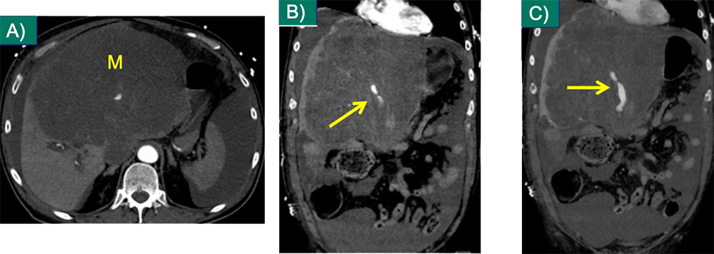
Pancreatic Metastasis

65-year-old woman with pancreatic mass due to metastatic melanoma. Axial image (A) and coronal MPR (B) from IV contrast enhanced CT show a hypovascular mass in the pancreatic body with ductal dilation in the distal body and tail. The metastasis mimics a primary pancreatic adenocarcinoma.

Pancreatic Metastasis


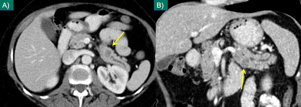
Small Bowel

56-year-old man with multifocal small bowel wall thickening due to metastatic melanoma involving both the jejunum (A) and ileum (B) on coronal IV contrast enhanced CT. Note that this appearance mimics lymphoma involving small bowel.

Small Bowel


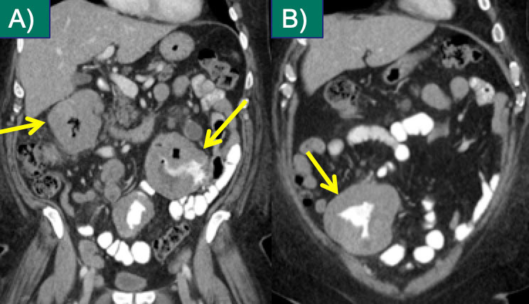
Small Bowel

78-year-old man with melanoma and nodular metastatic lesions in the small bowel wall on axial (C) and coronal (D) IV contrast enhanced CT. Small bowel should be inspected on axial and coronal planes to identify small metastases.

Small Bowel


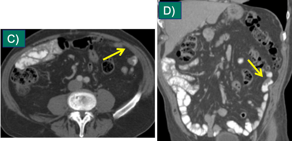
Mesenteric Metastasis

66-year-old man with melanoma and new mesenteric nodule on axial (A) and coronal (B) IV contrast enhanced CT. Coronal MPRs facilitate distinction of mesenteric metastases from bowel. Note intense FDG activity (C) on correlate PET image.

Mesenteric Metastasis


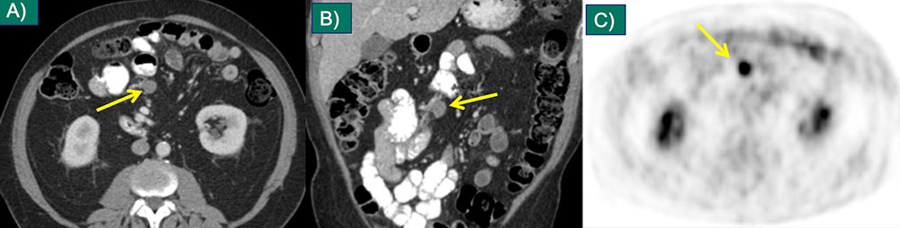
Perinephric Metastasis

55-year-old female with melanoma and small perinephric metastases on axial (A) and coronal (B) IV contrast-enhanced CT, which enlarged at 3 month follow up (C,D). (Same patient as slide 17)

Perinephric Metastasis


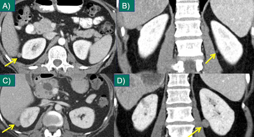
Uterine Metastasis

44-year-old female with melanoma and small uterine/periuterine nodule seen on axial IV contrast-enhanced CT (A). Intense FDG activity noted on follow-up PET/CT (B). No clear CT correlate (C) for a second focus of intense FDG activity in the uterus (D). PET/CT can identify some solid organ metastases not visualized with CT.

Uterine Metastasis


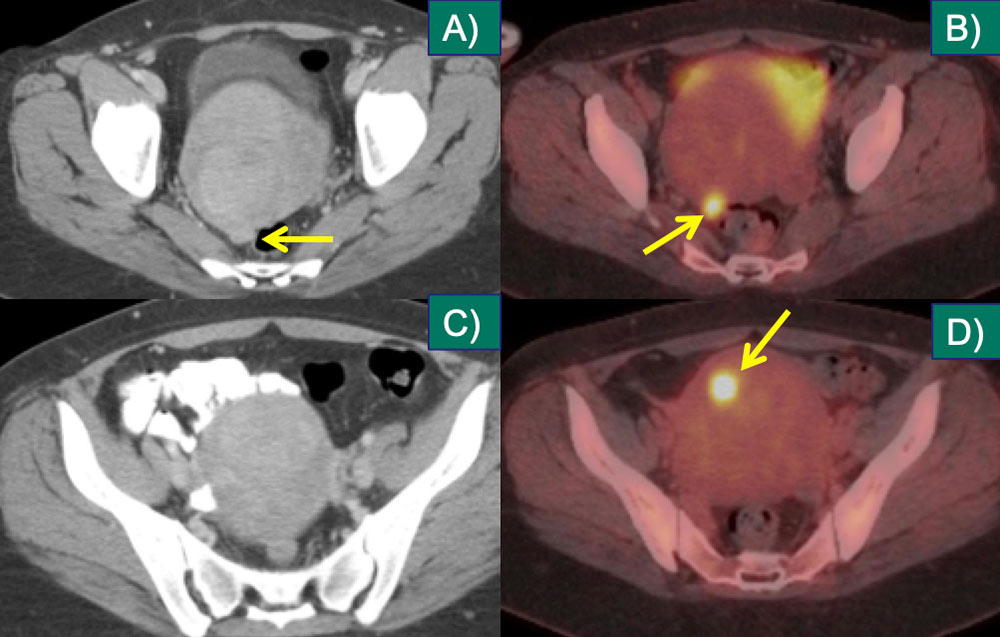
Cutaneous/Subcutaneous

56-year-old man with metastatic melanoma from right gluteal primary. Axial extended-field-of-view CT image demonstrates two skin metastases. Lower extremity, back, and head and neck melanoma metastasize to subcutaneous tissues.8

Cutaneous/Subcutaneous


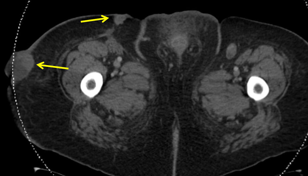
Isolated Subcutaneous Metastasis

51-year old woman with melanoma and small subcutaneous nodule that increased from 4 mm (A) to 8 mm (B). Note FDG avidity on PET/CT (C). This was the only site of metastatic disease. Careful inspection of the subcutaneous tissues with comparison to prior exams is imperative; subcutaneous metastases are often small at presentation.

Isolated Subcutaneous Metastasis


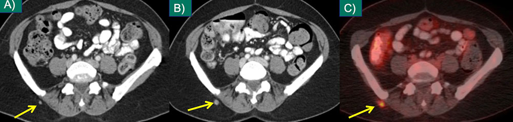
Complications of Melanoma Mets

57-year-old male with metastatic (M) to liver lesion on axial CT (A) and active hemorrhage on IV contrast enhanced MDCT arterial (B) and venous (C) phase coronal MPRS.
  • Melanoma metastases have a propensity for hemorrhage, and may present with significant GI hemorrhage10
  • Melanoma metastatic to the small bowel may result in obstruction or serve as a lead point for intussusception
Complications of Melanoma Mets


On the Treatment Horizon

Adoptive cell transfer:
  • Isolation, expansion, and reinfusion of tumor infiltrating lymphocytes found in sites of disease (or modification of peripherally isolated T cells to express a T cell receptor that targets melanoma)
  • Response rates from 34-72%25
Therapy for oligometastatic disease:
  • Surgical metastasectomy improves median and 5-year survival in M1a (skin, subcutaneous, distant nodes) and M1b/M1c (visceral or osseous) disease26
  • Resection of pulmonary metastases with no extrathoracic disease also improves survival, even in patients with >5 year disease free interval27
  • Resection safe during BRAF-inhibitor therapy28
  • Stereotactic radiotherapy may be an alternative to surgery; for example, for pulmonary mets29


Immunotherapy Complications

Axial (A,B), sagittal (C), and coronal (D) IV and oral contrast enhanced CT demonstrating diffuse colonic wall thickening in a 68-year-old female on ipilimumab for therapy of metastatic melanoma. Severe colitis was found on subsequent colonoscopy: pathologic changes consistent with ipilimumab-related disease.

Immunotherapy Complications


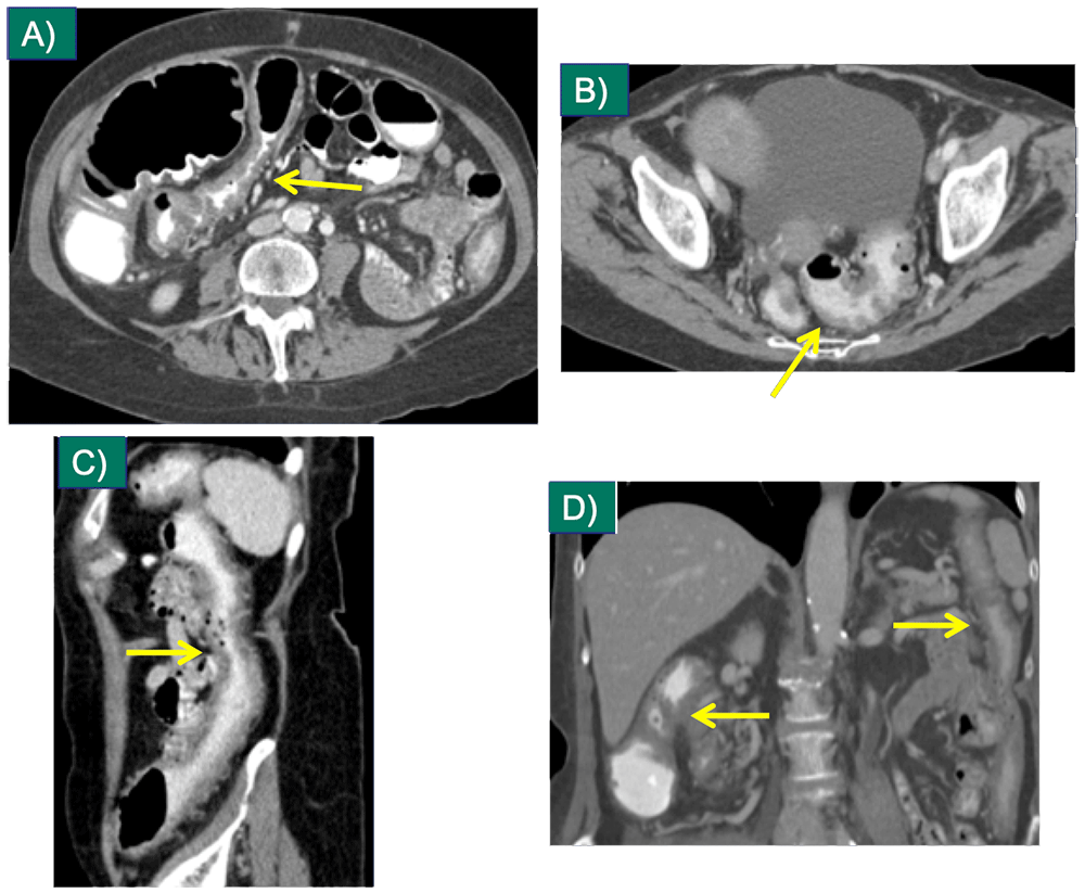
Immunotherapy Complications

Axial non-contrast CT of the chest in lung windows (A,B) demonstrating consolidation in the left lung, consistent with pneumonitis in a patient who had received 4 doses of ipilimumab for therapy of melanoma.

Immunotherapy Complications


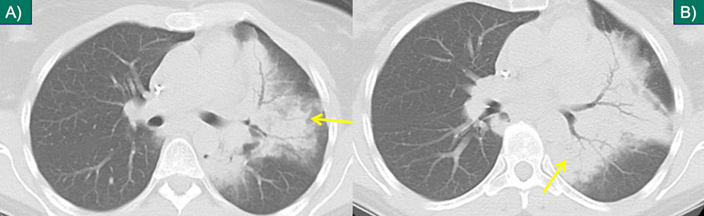
Immunotherapy Complications

Axial (A-C) IV contrast-enhanced CT in a 68-year-old male with metastatic melanoma on ipilimumab presenting with abdominal pain and elevated lipase, with pancreatic edema and adjacent inflammatory stranding, compatible with acute pancreatitis.
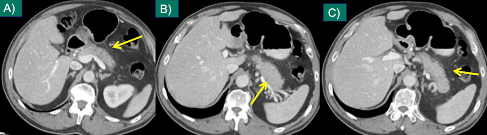
Immunotherapy Complications


References

  1. Siegel R, et al. Cancer statistics, 2014. CA Cancer J Clin 2014; 64:9.
  2. Guy GP Jrr, et al. Vital signs: melanoma incidence and mortality trends and projections - United States, 1982-2030. MMWR Morb Mortal Wkly Rep 2015; 64:591.
  3. Ekwueme DU et al. The health burden and economic costs of cutaneous melanoma mortality by race/ethnicity—United States, 2000 to 2006. J Am Acad Dermatol 2011; 65:S133–S143.
  4. Guy GP, et al. Prevalence and costs of skin cancer treatment in the US, 2002-2006 and 2007-2011. Am J Prev Med 2015; 48:183–7.
  5. Ferlay J, et al. Estimates of worldwide burden of cancer in 2008: GLOBOCAN 2008. Int J Cancer 2010; 127:2893.
  6. Parkin DM, Pisani P, Ferlay J. Estimates of the worldwide incidence of 25 major cancers in 1990. Int J Cancer 1999; 80:827.
  7. Patel JK, et al. Metastatic pattern of malignant melanoma. A study of 216 autopsy cases. Am J Surg 1978; 135: 807–810.
  8. Trout AT et al. Melanoma metastases in the abdomen and pelvis: Frequency and patterns of spread. WJR 2013 5(2): 25-32.
  9. Bender GN, et al. Malignant melanoma: patterns of metastasis to the small bowel, reliability of imaging studies, and clinical relevance. Am J Gastroenterol 2001; 96(8):2392-2400.
  10. King DM. Imaging of Metastatic Melanoma. J HK Coll Radiol 2004 7:66-69.
  11. Winkler N, Rezvani M, Heilbrun M, Shaaban A. Utility of dual phase liver CT for metastatic melanoma staging and surveillance. Eur J Radiol. 2013 Dec;82(12):2189-93.
  12. Haider MA, Amitai MM, Rappaport DC, O'Malley ME, Hanbidge AE, Redston M, Lockwood GA, Gallinger S. Multi-detector row helical CT in preoperative assessment of small (< or = 1.5 cm) liver metastases: is thinner collimation better? Radiology. 2002 Oct;225(1):137-42
  13. Khan ZS, Huth J, Kapur P, Huerta S. Indications and recommended approach for surgical intervention of metastatic disease to the gallbladder. World J Surg Oncol. 2010 Sep 10;8:80.
  14. Perdona S, et al. A case of renal melanoma metastasis: description of clinico-pathological features. Arch Ital Urol Androl 2007; 79(4):161-163.
  15. Ozyuncu N et al. Cardiac metastasis of malignant melanoma: a rare case of complete atrioventricular block. Europace 2006; 8:545-548.
  16. Meunier R, Pareek G, Amin A. Metastasis of malignant melanoma to urinary bladder: A case report and review of the literature. Case Rep Path 2015; 2015: 6 pages, Article ID 173870.
  17. Liang K, et al. Metastatic malignant melanoma of the gastrointestinal tract. Mayo Clin Proc 2008; 81(4):511-516.
  18. Reccia I, et al. An uncommon presentation of metastatic melanoma: a case report. Medicine 2015; 94(7):1-4.
  19. Xiong Y, et al. Pelvis metastasis from primary choroidal melanoma: a case report. OncoTargets Ther 2014; 7:2107-2110.
  20. Junquels C, et al. An unusual digestive occlusion [French]. Rev Med Brux 2015 ; 36(1):42-44.
  21. Postow MA, et al. Nivolumab and ipilimumab versus ipilimumab in untreated melanoma. N Engl J Med 2015; 372:2006-2017.
  22. Maio M, et al. Five-year survival rates for treatment-naïve patients with advanced melanoma who received ipilimumab plus dacarbazine in a phase III trial. J Clin Oncol 2015; 33:1191-1196.
  23. Wyluda EJ, et al. Durable complete responses off all treatment in patients with metastatic malignant melanoma after sequential immunotherapy followed by a finite course of BRAF inhibitor therapy. Cancer Biol Ther 2015; 16(5):662-670.
  24. Johnson DB, et al. Combined BRAF (Dabrafenib) and MEK inhibition (trametinib) in patients with BRAFV600-mutant melanoma experiencing progression with single-agent BRAF inhibitor. J Clin Oncol 2014; 32:3697-3704.
  25. Finn L, Markovic SN, Joseph RW. Therapy for metastatic melanoma: the past, present, and future. BMC Medicine 2012; 10:23.
  26. Wasif N, Bagaria SP, Ray P, Morton DL. Does metastasectomy improve survival in patients with Stage IV melanoma? A cancer registry analysis of outcomes. J Surg Oncol. 2011;104:111–5.
  27. Petersen RP, Hanish SI, Haney JC, et al. Improved survival with pulmonary metastasectomy: an analysis of 1720 patients with pulmonary metastatic melanoma. J Thorac Cardiovasc Surg. 2007;133:104–10.
  28. He M, et al. Post-Operative Survival Following Metastasectomy for Patients Receiving BRAF Inhibitor Therapy is Associated With Duration of Pre-Operative Treatment and Elective Indication. J Surg Onc. 2015;111:980–984.
  29. Singh D, et al. Local control rates with five-fraction stereotactic body radiotherapy for oligometastatic cancer to the lung. J Thorac Dis 2014;6(4):369-374.
  30. Tirumani SH, et al. Radiographic Profiling of Immune-Related Adverse Events in Advanced Melanoma Patients Treated with Ipilimumab. Cancer Immunol Res 2015 Jun 22. [Epub ahead of print]. Accessed 7/19/2015.
  31. Raad RA, et al. Imilimumab-induced hepatitis on 18F-FDG PET/CT in a patient with malignant melanoma. Clin Nucl Med 2015; 40:258-259.
  32. Bacanovic S, et al. Ipilimumab-induced adrenalitis: a possible pitfall in 18F-FDG PET/CT. Clin Nucl Med 2015 Jul 7 [Epub ahead of print]. Accessed 7/19/2015.
  33. Yu C, Chopra IJ, Ha E. A novel melanoma therapy stirs up a storm: ipilimumab-induced thyrotoxicosis. Endocrinol Diab Metab Case Reports 2015 ;2015:140092.
  34. Fishman EK, et al. CT of malignant melanoma in the chest, abdomen, and musculoskeletal system. RadioGraphics 1990; 10:603-620.
  35. Nishino M, et al. Response assessment in metastatic melanoma treated with ipilimumab and bevacizumab: CT tumor size and density as markers for response and outcome. J ImmunoTherapy Cancer 2014, 2:40.
  36. Shrot S, et al. CT halo sign as an imaging marker for response to adoptive cell therapy in metastatic melanoma with pulmonary metastases. Eur Radiol 2014; 24:1251–1256.
  37. Beaino W, Nedrow JR, Anderson CJ. Evaluation of 68Ga- and 177Lu-DOTA-PEG4‑LLP2A for VLA-4-targeted PET imaging and treatment of metastatic melanoma. Mol Pharmaceutics 2015; 12: 1929−1938.
  38. Carling U, et al. Transarterial chemoembolization of liver metastases from uveal melanoma using irinotecan-loaded beads: treatment tesponse and complications. Cardiovasc Intervent Radiol 2015 Apr 2. [Epub ahead of print] . Accessed 7/19/2015.
  39. Schelhorn J, et al. A single-center experience in radioembolization as salvage therapy of hepatic metastases of uveal melanoma. Acta Radiologica Open 2015; 4(4): 1–7.
  40. Pfluger T, Melzer HI, Schneider V, La Fougere C, Coppenrath E, Berking C, Bartenstein P, Weiss M. PET/CT in malignant melanoma: contrast-enhanced CT versus plain low-dose CT. Eur J Nucl Med Mol Imaging. 2011 May;38(5):822-31.

Privacy Policy

Copyright © 2024 The Johns Hopkins University, The Johns Hopkins Hospital, and The Johns Hopkins Health System Corporation. All rights reserved.