google ads

CT of Uncomplicated and Complicated Gastric Volvulus: Unraveling the Imaging Findings with Volumetric Dataset Interpretations

CT of Uncomplicated and Complicated Gastric Volvulus: Unraveling the Imaging Findings with Volumetric Dataset Interpretations


Gastric Volvulus

  • Defined as pathologic rotation of the stomach.
  • Subtypes depends on which axis the rotation occurs.
    • Longitudinal axis = Organoaxial (2/3 of cases in adults)[1]
    • Transverse axis = Mesenteroaxial (more common type in pediatric patients)
    • Less common is a combined type


Gastric Volvulus

  • Primary (sub diaphragmatic) Volvulus (1/3 of cases)[2]
  • Majority occur secondarily due to a combination of a predisposing factor and laxity of the suspensory ligaments
    • Paraesophageal Hiatus Hernia
    • Traumatic diaphragmatic hernia
    • Eventration / elevation of diaphragm


Organoaxial Volvulus

  • Stomach rotates along its long axis
  • Greater curvature displaced superiorly (dashed yellow line)
  • Less curvature located more caudally(solid red line)
  • Fundus rotates posteroinferiorly (F)
  • Antrum rotates anterosuperiorly (A)
Organoaxial Volvulus


Mesenteroaxial Volvulus

  • Less common
  • Stomach rotates along short axis
  • Antrum displaced above gastroesophageal junction
Mesenteroaxial Volvulus


Acute Gastric Volvulus

  • Signs and symptoms depend on the chronicity, the degree of rotation, degree of obstruction and location relative to diaphragm.
    • Primary volvulus (subdiaphragmatic) typically have abdominal pain, distention and severe vomiting.
    • Secondary volvulus typically have similar presentation often with radiating chest pain.
  • Rate of transformation of asymptomatic gastric volvulus to acute volvulus is unknown.
  • Borchardt in 1904 described a triad of symptoms[4]:
    • Severe epigastric pain and distention.
    • Vomiting followed by violent nonproductive retching.
    • Difficulty to pass a nasogastric tube.


Complications of Acute Volvulus

  • Mortality reported as high as 30-50% [5]
    • With prompt recognition and intervention, mortality rate drops (as low as 16%)
  • Strangulation: 5-28% of cases [3]
    • Due to rich blood supply of the stomach
    • More common with organoaxial rotation
  • Other possible reported complications include:
    • Ulceration
    • Perforation
    • Hemorrhage
    • Necrosis
    • Pancreatic, splenic or omental ischemia (secondary to disruption of adjacent vessels)


Imaging of Gastric Volvulus

  • Plain film
    • Insensitive. Can demonstrate hiatal hernia or suggest volvulus depending on symptoms, particularly with gastric air fluid level.
  • Upper GI / Barium swallow fluoroscopic study
    • Good test to characterize hiatal hernia and chronic gastric volvulus
  • MDCT
    • Test of choice in patients with acute abdominal symptoms


MDCT of Complicated Volvulus

  • Narrow collimation detectors for high quality multiplanar reformations (MPRs)
  • Dual phase (arterial and venous) IV contrast and PO water ideally
    • Arterial phase for preoperative vascular mapping
    • Evaluation of gastric wall for ischemia improved by use of IV contrast against neutral oral contrast in gastric lumen
    • Obstruction can be identified without positive oral contrast
  • If IV contrast can not be administered- positive oral contrast
  • Coronal and sagittal MPRs are critical for defining configuration and identifying complications


MDCT of Complicated Volvulus

  • Majority of cases are associated with a predisposing factor
    • Most commonly large paraesophageal hernia
    • Post traumatic diaphragmatic hernia
    • Elevated diaphragm
  • More often the rotation is less than 180 degrees, resulting in incomplete or partial volvulus
    • Often lack of symptoms or secondary findings of obstruction on imaging


MDCT of Complicated Volvulus

  • Gastric outlet obstruction can occur when twist is greater than 180 degrees [1]
    • Severe gastric distention
    • Air – fluid level
    • Gastric transition point
      • Most often an antropyloric narrowing
      • Commonly at the esophageal diaphragmatic hiatus


MDCT of Complicated Volvulus

  • Signs of strangulation / ischemia
    • Gastric wall thickening
    • Mucosal hypoenhancement
    • Perigastric fluid
    • Gastric pneumatosis
  • Pneumoperitoneum (secondary to perforation)
  • Pancreatic, splenic or omental ischemia (secondary to disruption of adjacent vessels)


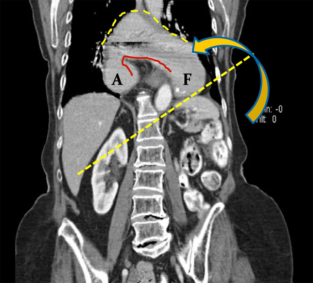
Case 1. Organoaxial

Coronal MPR from IV contrast enhanced MDCT shows uncomplicated organoaxial gastric volvulus, secondary to a complete hiatal hernia.
A= antrum
F= fundus
Case 1. Organoaxial


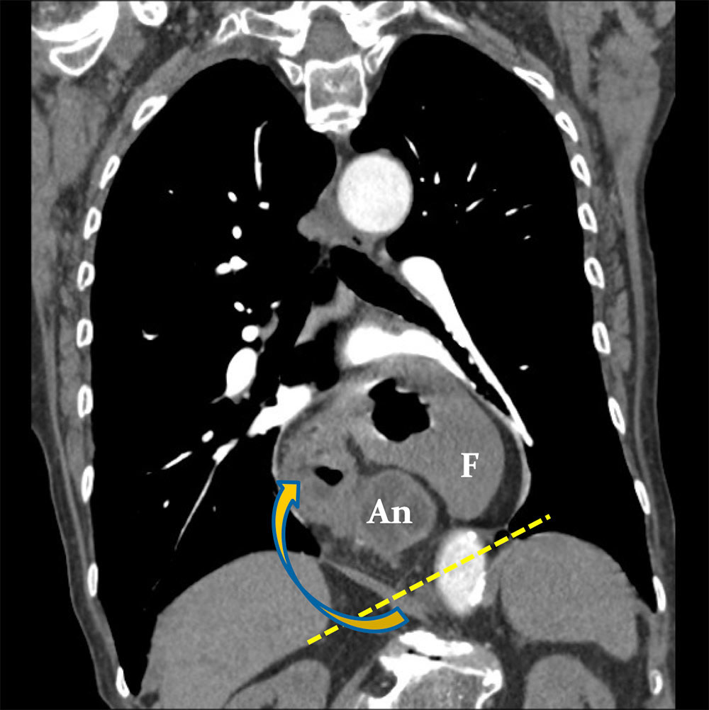
Case 2. Mesenteroaxial

Coronal MPR from IV contrast enhanced MDCT shows uncomplicated chronic mesenteroaxial gastric volvulus, secondary to large diaphragmatic hernia.
An= antrum
F= fundus

Case 2. Mesenteroaxial


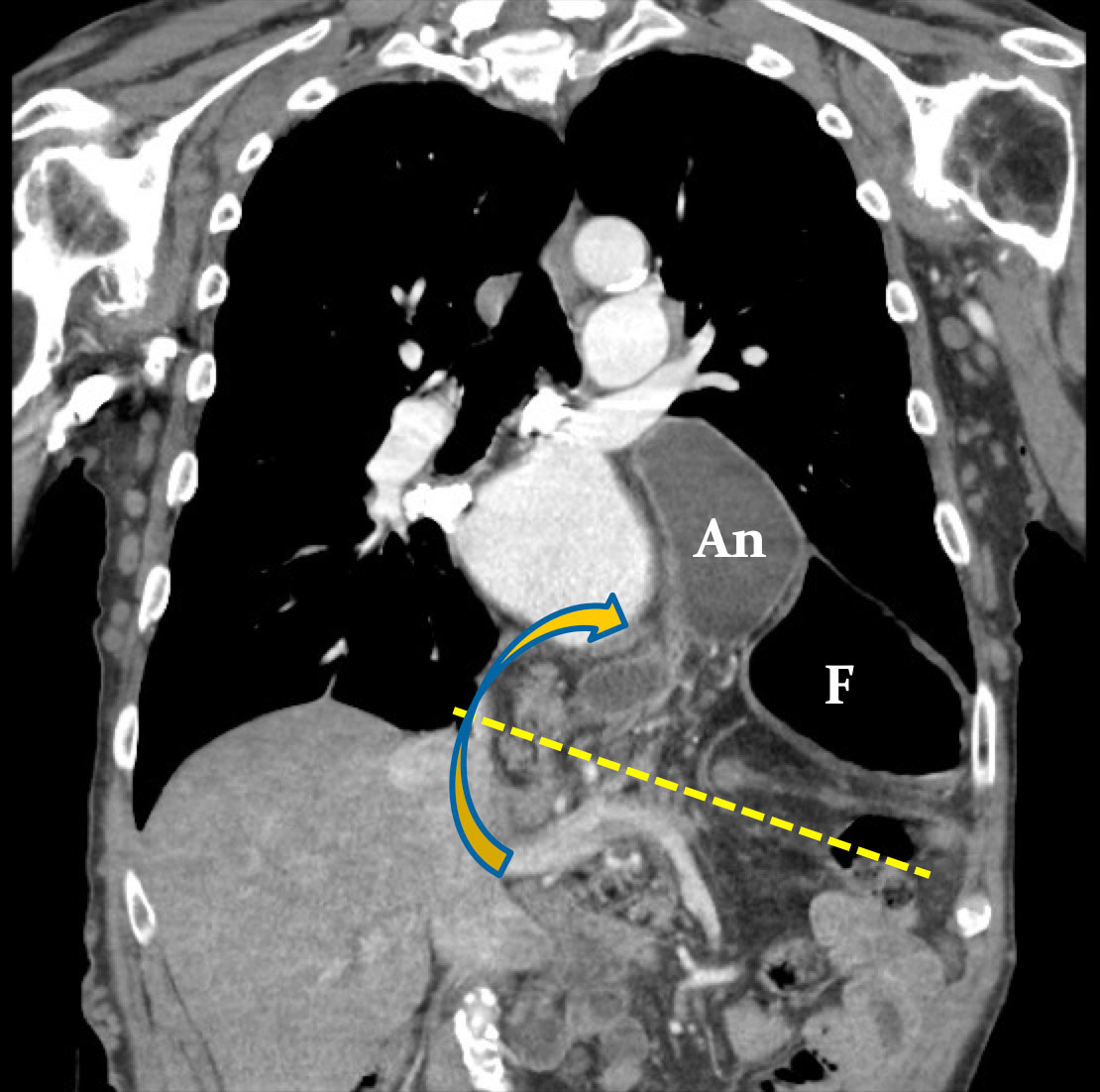
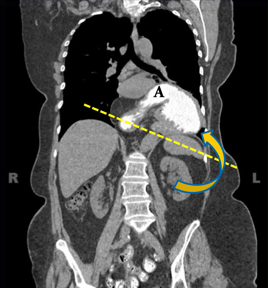
Case 3. Organoaxial

Coronal MPR from noncontrast MDCT shows uncomplicated organoaxial gastric volvulus, secondary to a large hiatal hernia.

Case 3. Organoaxial


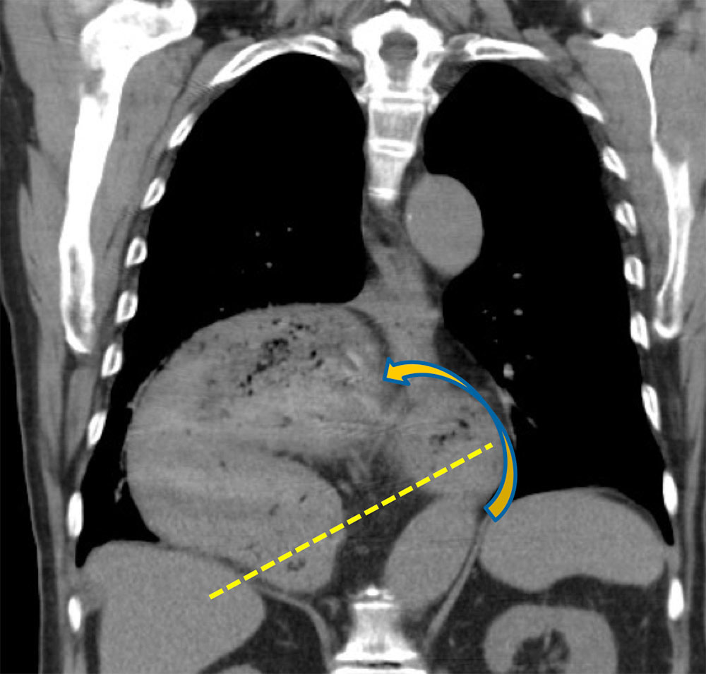
Case 4. Mesenteroaxial

Coronal MPR from IV contrast enhanced MDCT (A) of chronic uncomplicated mesenteroaxial gastric volvulus, secondary to large diaphragmatic hernia. (B) Subsequent Upper GI examination.

Case 4. Mesenteroaxial


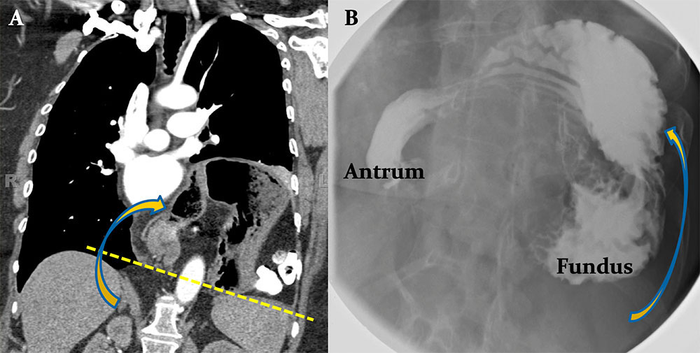
Case 5. Organoaxial

Coronal MPR (A) and axial image (B) from oral and IV contrast enhanced MDCT shows uncomplicated organoaxial gastric volvulus, secondary to a large hiatal hernia.

Case 5. Organoaxial


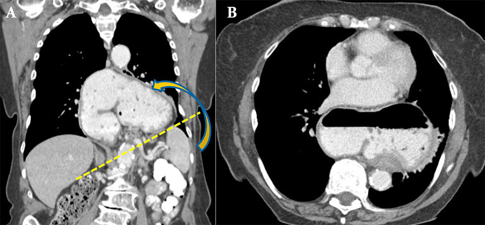
Case 6. Mesenteroaxial

Coronal (A) and sagittal (B) MPRs from oral contrast enhanced MDCT show uncomplicated mesenteroaxial gastric volvulus, secondary to large diaphragmatic hernia. Notice the antrum (An) lying above the fundus (F).

Case 6. Mesenteroaxial


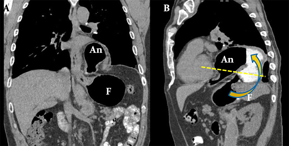
Case 7. Chronic Mixed Type Volvulus

Coronal (A) and sagittal (B) MPRs from oral contrast enhanced Chronic gastric volvulus of the mixed / combined type. Sagittal (A) and coronal (B) MPRs from IV contrast enhanced MDCT show complex twisting and turning of the stomach, but not along any particular axis. The cause is secondary to eventration and elevation of the posterior diaphragm.

Case 7. Chronic Mixed Type Volvulus


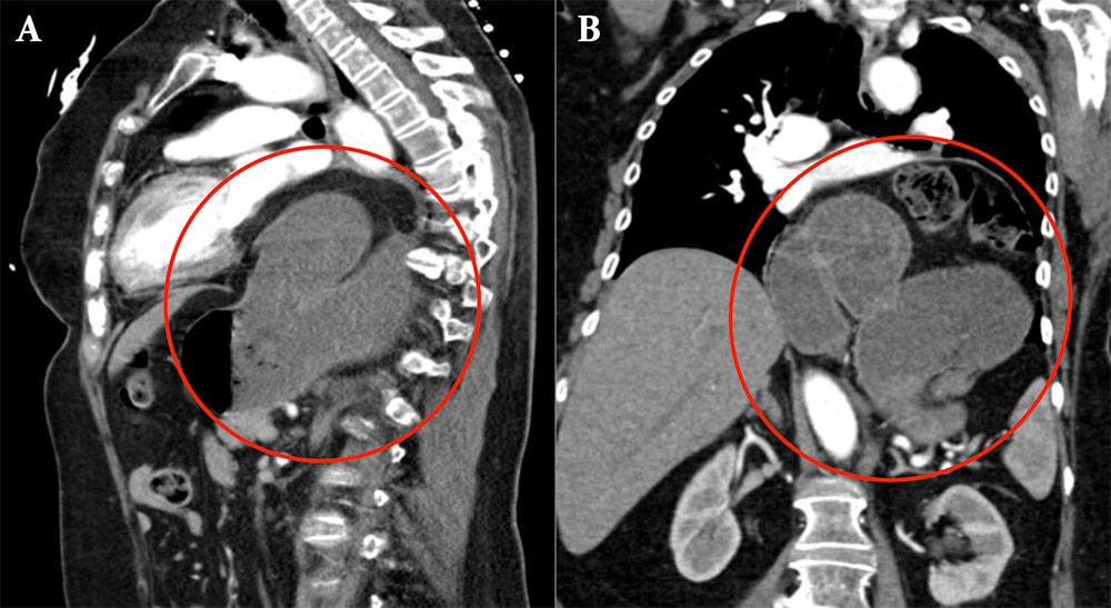
Case 8. Mixed Type Volvulus

Axial image (A) and coronal MPR (B) from IV and oral contrast enhanced MDCT demonstrate complex turning of the stomach (circle), but not along any particular axis, compatible with a mixed type volvulus.

Case 8. Mixed Type Volvulus


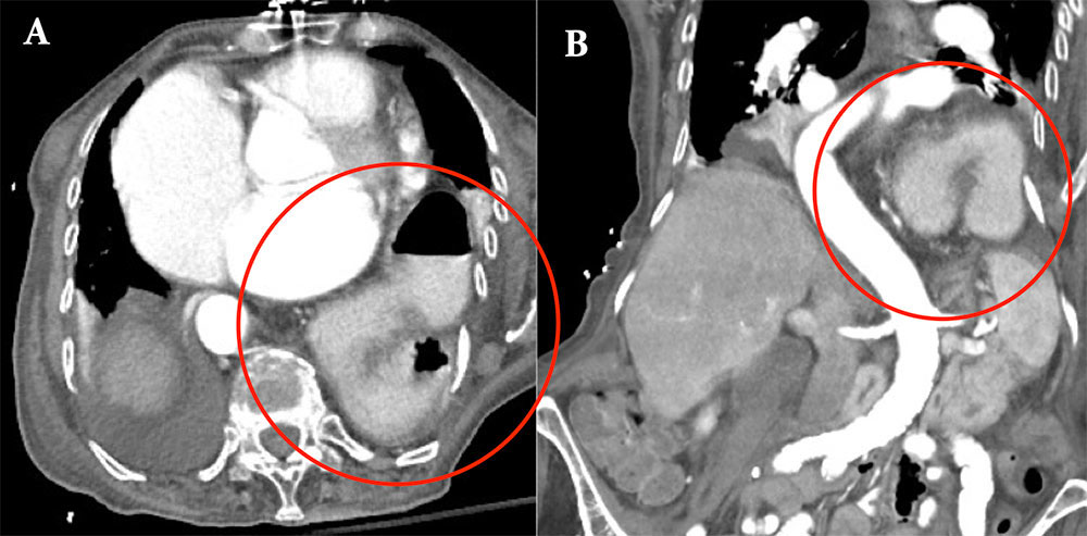
Case 9. Mixed Type Volvulus

Axial image (A) and sagittal MPR (B) from IV contrast enhanced MDCT in a patient with recurrent mild abdominal symptoms demonstrate an uncomplicated mixed type volvulus (circle).

Case 9. Mixed Type Volvulus


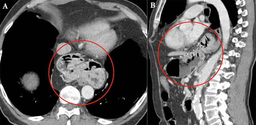
Case 10. Mixed Type Volvulus

Coronal 3D volume rendering (A), coronal MPR (B) and axial image (C) from IV contrast enhanced MDCT demonstrate complex twisting of the stomach, but not along any particular axis, compatible with a mixed type volvulus.

Case 10. Mixed Type Volvulus


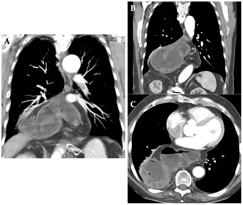
Case 11. Volvulus with Obstruction

Coronal MPR (A) and axial image (B) from oral and IV contrast enhanced MDCT show organoaxial volvulus with outlet obstruction at the antropyloric region, narrowed by the distended body (arrow). Note the gastric distention with ingested material and air fluid level (*). No additional findings to suggest ischemia.

Case 11. Volvulus with Obstruction


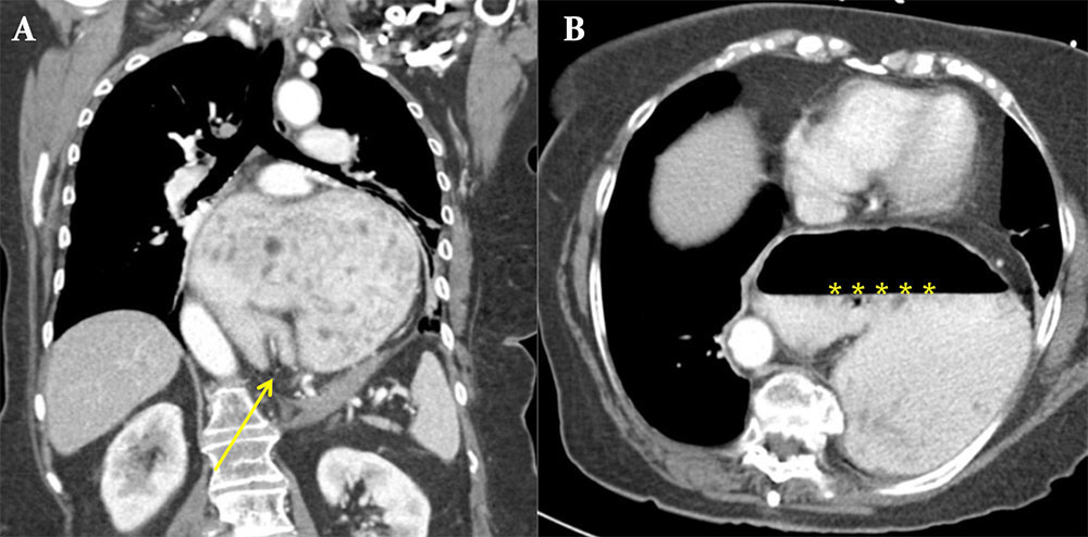
Case 12. Volvulus with Obstruction

Coronal MPR (A), axial image (B) and coronal 3D volume rendering (C) from IV contrast enhanced MDCT show a large hiatal hernia with gastric volvulus and high grade outlet obstruction at the level of the antropyloric region (arrows). Again seen is gastric distention with air fluid level (*).

Case 12. Volvulus with Obstruction


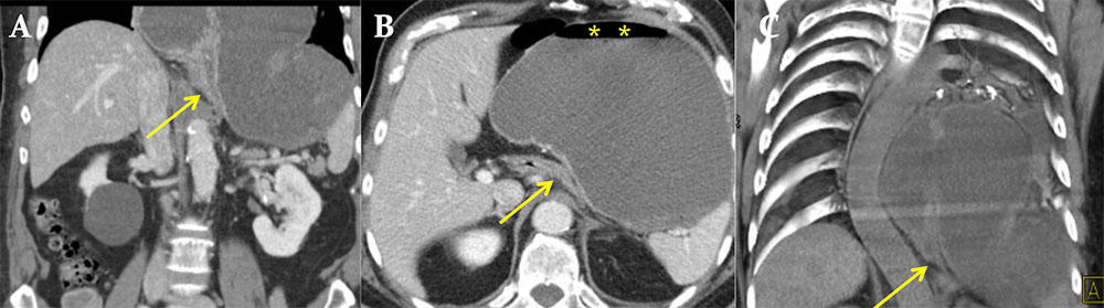
Case 13. Volvulus with Obstruction

Coronal MPR from IV contrast enhanced MD CT baseline study (A) in a patient with chronic organoaxial gastric volvulus. Chest radiograph (B), axial oral and IV contrast enhanced MDCT (C,D) when patient developed acute outlet obstruction at the antropyloric region traversing through the esophageal hiatus (arrow). Gastric distention with air fluid level (*). An = antrum, F = fundus.

Case 13. Volvulus with Obstruction


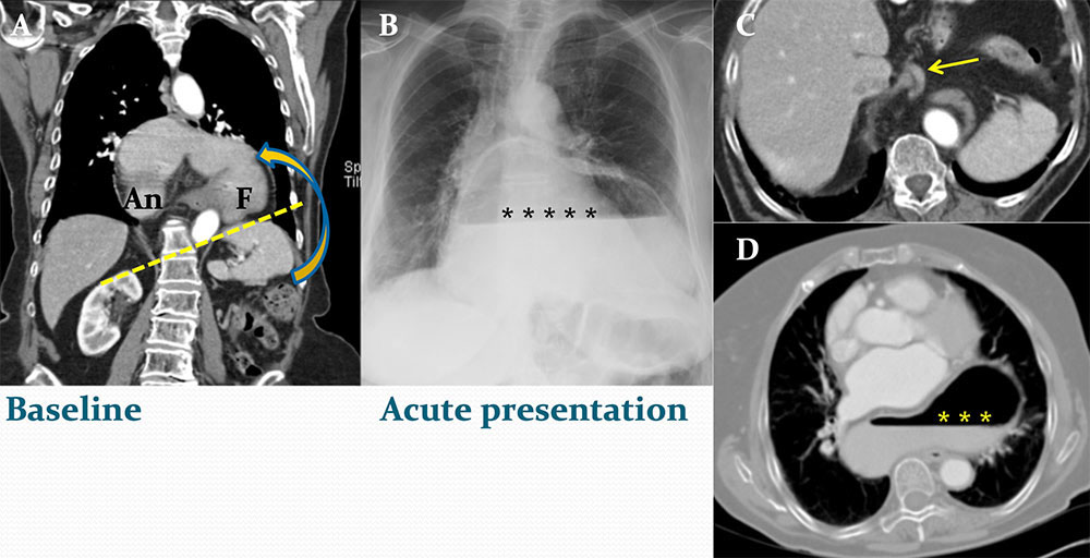
Case 14. Volvulus with Ascites

Coronal MPR (A) and axial image (B) from IV contrast enhanced MDCT show organoaxial gastric volvulus with small amount perigastric ascites (arrows). There is no obstruction nor gastric wall thickening. Adjacent fluid as an isolated secondary finding is non specific; nonetheless, it is noteworthy and important to inform clinicians to correlate with symptoms, as intermittent strangulation is possible.

Case 14. Volvulus with Ascites


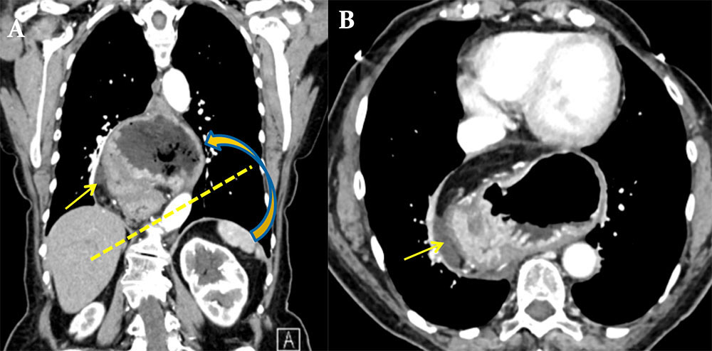
Case 15. Volvulus with Obstruction and Strangulation

Baseline coronal MPR (A) from oral and IV contrast enhanced MDCT demonstrates large hiatal hernia with chronic volvulus (circle). Patient presented at follow up with acute abdominal pain. Axial image (B) and coronal MPR (C) from oral and IV contrast enhanced CT demonstrates luminal distention and gastric wall thickening (arrows), an indicator of strangulation in the setting of obstruction.

Case 15. Volvulus with Obstruction and Strangulation


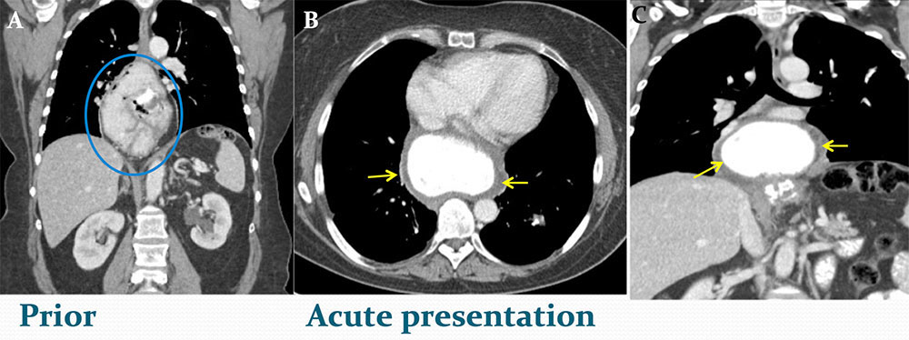
Case 16: Volvulus with Gastric Ischemia

Axial image (A) and coronal MPR (B) from oral contrast enhanced MDCT show organoaxial volvulus with outlet obstruction and signs of ischemia, as evidenced by bowel walk thickening (solid arrow) and perigastric fluid (dashed arrow). No gastric emphysema.
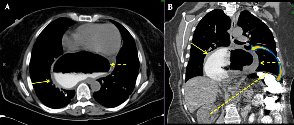
Case 16: Volvulus with Gastric Ischemia


Treatment

  • Chronic Gastric Volvulus
    • Conservative treatment with watchful observation.
    • 64% of patients without correction will experience recurrent symptoms [7].
  • Acute Gastric volvulus is a surgical emergency
    • Surgical gastric reduction
    • Gastric fixation (gastropexy)
    • Repair of any associated diaphragmatic hernia with addition of fundoplication [8]


Summary

  • Look closely with large hiatal hernias or diaphragmatic abnormalities for possibility of underlying gastric volvulus.
  • Evaluation of the patient with gastric volvulus requires dataset interpretation with axial images and multiplanar reconstructions.
  • Recognize signs of complicated gastric volvulus such as gastric outlet obstruction, strangulation or ischemia, and promptly communicate to ordering clinicians.


References

  1. Peterson CM, Anderson JS, Hara AK, Carenza JW, Menias CO(2009) Volvulus of the gastrointestinal tract: appearances at multimodality imaging. Radiographics 29:1281–1293.
  2. M.H. Sleisenger, J.S. Fordtran (Eds.), Gastrointestinal disease: pathophysiology, diagnosis, management (5th edn.), WB Saunders, Philadelphia (1993), pp. 481–483.
  3. J.A. Wasselle, J. Norman. Acute gastric volvulus: pathogenesis, diagnosis, and treatment. Am J Gastroenterol, 88 (1993), pp. 1780–1784.
  4. M. Borchardt. Zun pathologie and therapie des magnevolvulus. Arch Klin Chir, 74 (1904), pp. 243–248.
  5. R.J. Smith. Volvulus of the stomach. JAMA, 75 (1983), pp. 393–396.
  6. Millet I, Orliac C, Alili C, Guillon F, Taourel P. Computed Tomography Findings of Acute Gastric Volvulus. Eur Radiol. 2014 Oct 4.
  7. Hsu Y, Perng C, Chen C, Tsai J, Lin H. Conservative Management of Chronic Gastric Volvulus: 44 cases over 5 years. World J Gastroeneterol, 2010 Sept 7; 16(33):4200-4205.
  8. Gourgiotis S, Vougas V, Germanos S, Baratsis S. Acute Gastric Volvulus: Diagnosis and Management over 10 Years. Dig Surg, 2006; 23:169-172.
Acknowledgements:
  • Hazem Hawasli, M.D.
  • Pamela T. Johnson, M.D
  • Karen M. Horton, M.D.
  • Elliot K. Fishman, M.D.

Privacy Policy

Copyright © 2024 The Johns Hopkins University, The Johns Hopkins Hospital, and The Johns Hopkins Health System Corporation. All rights reserved.