A Beginner's Guide to Cinematic Rendering in Clinical Practice: What You Need to Know
A Beginner's Guide to Cinematic Rendering in Clinical Practice: What You Need to Know Linda C. Chu, MD, Steven P. Rowe, MD PhD, and Elliot K. Fishman, MD The Russell H. Morgan Department of Radiology and Radiological Science, Johns Hopkins University, Baltimore, MD |
Cinematic Rendering (CR)
|
Cinematic Rendering (CR) CR uses global illumination model that takes direct and indirect illumination into account to generate photorealistic images Volume Rendering (VR)
|
Why We Do It? 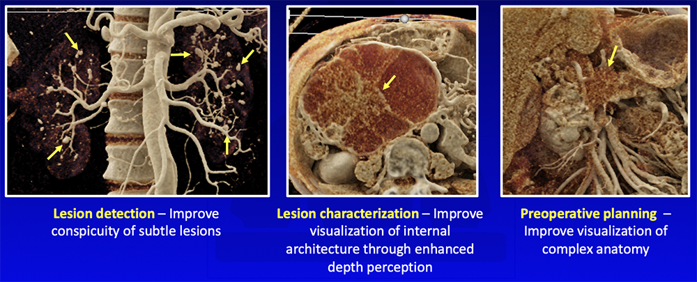 |
When We Do It?
|
Cardiovascular Applications
|
Cardiovascular – Congenital Anomalies 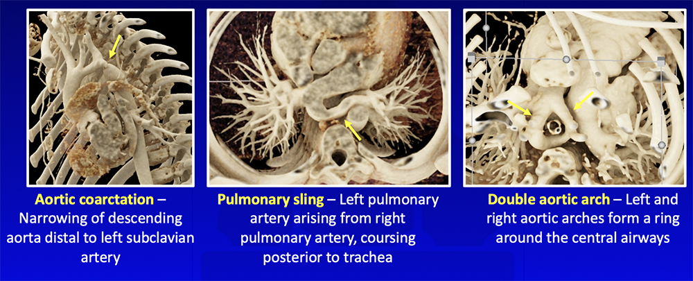 |
Cardiovascular – Coronary Artery Anomalies 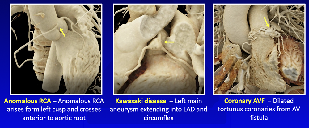 |
Cardiovascular – Complex Vascular Anatomy 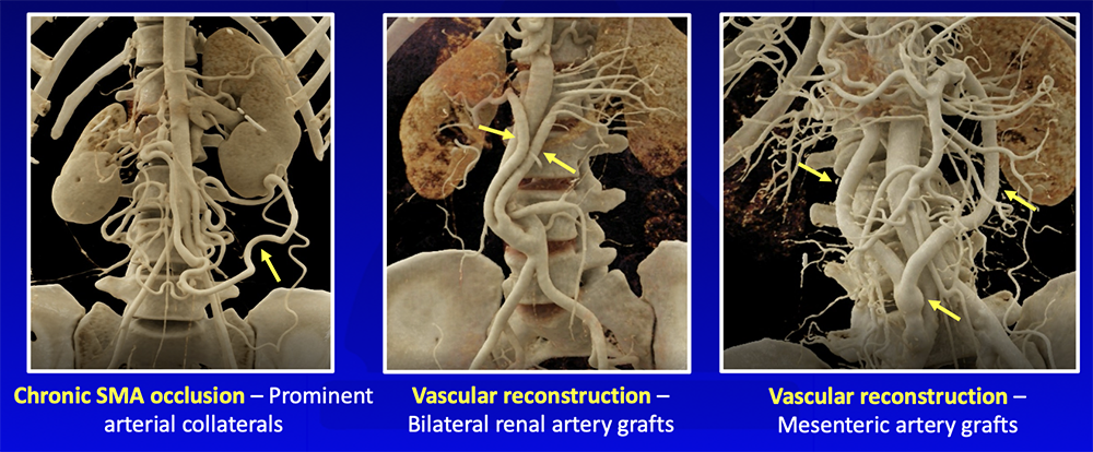 |
Cardiovascular – Vasculitis 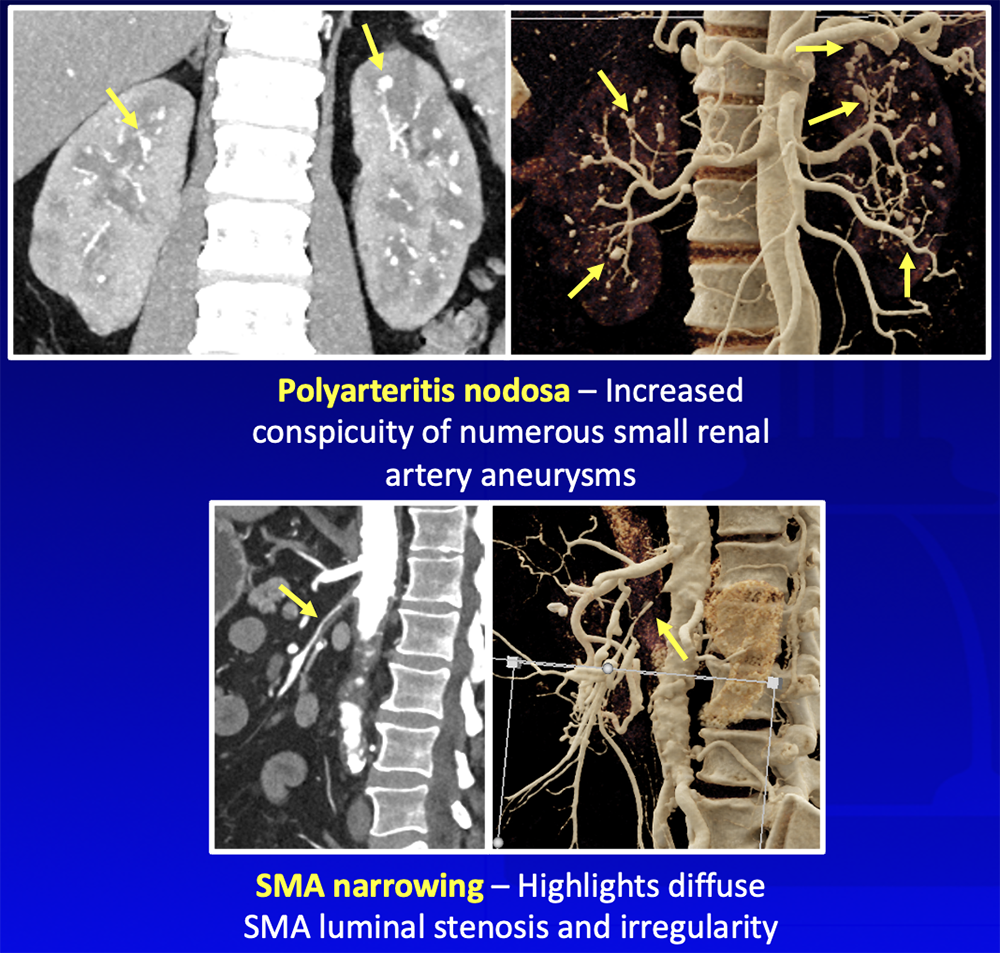 |
Cardiovascular – Preoperative Planning 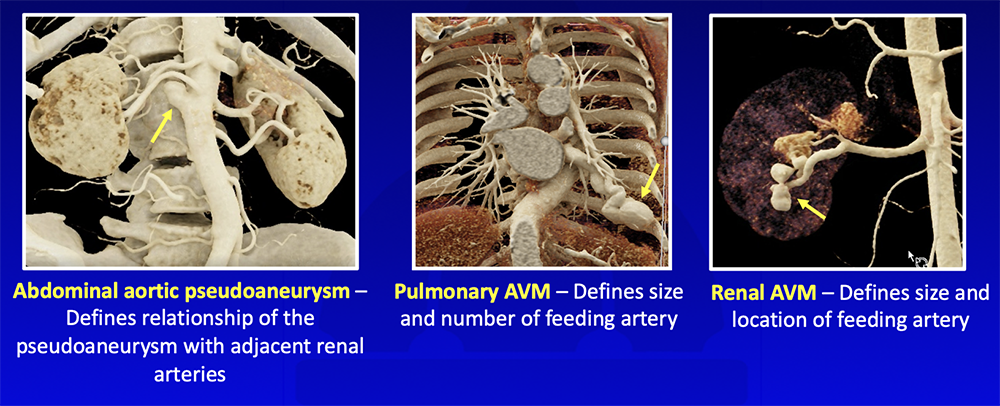 |
Chest Applications
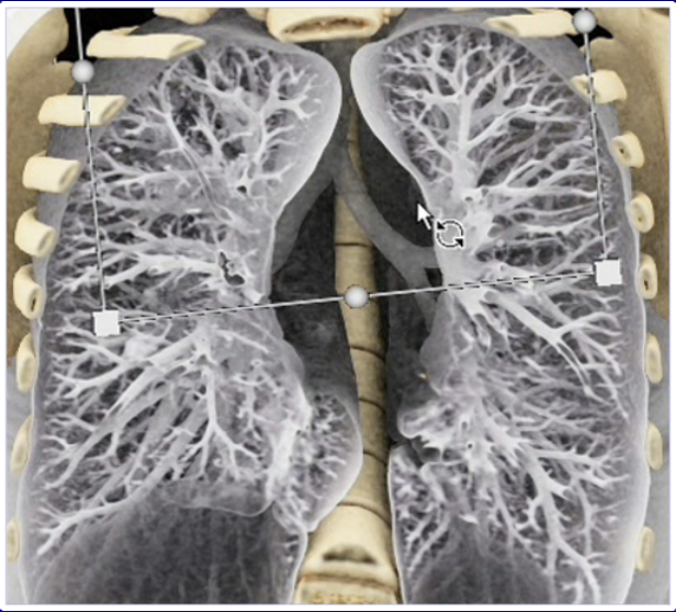 |
Chest Applications Recht HS et al. Diag Interv Imaging. 2022;103(2):123-124. 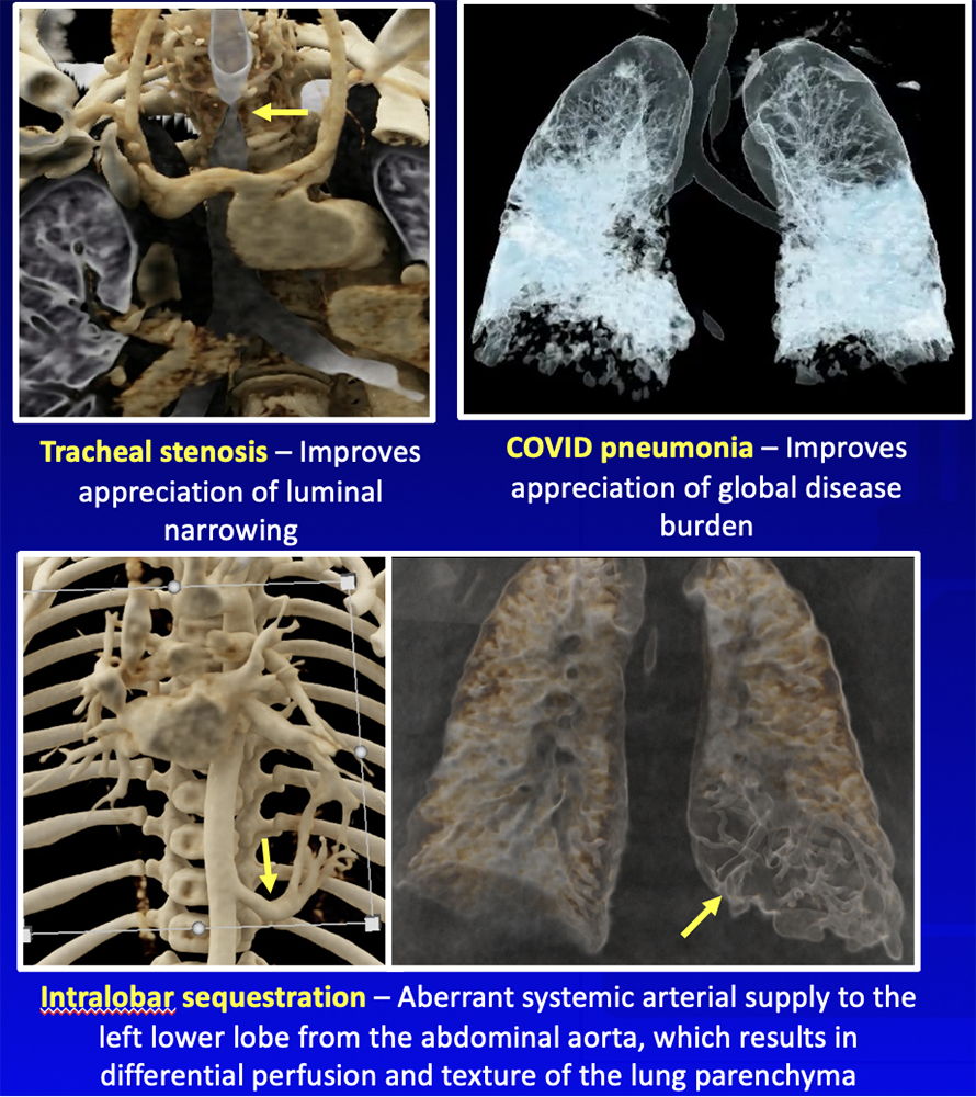 |
Trauma/Musculoskeletal Applications
|
Trauma/Musculoskeletal 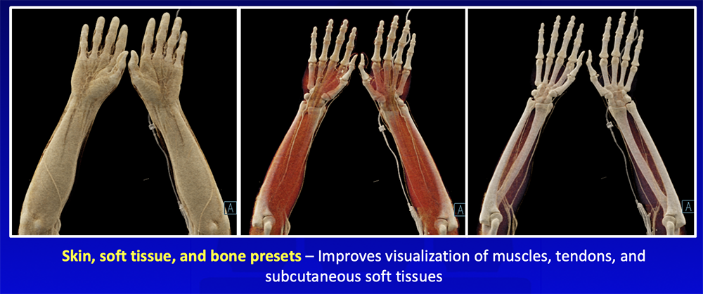 |
Trauma – Musculoskeletal Injury 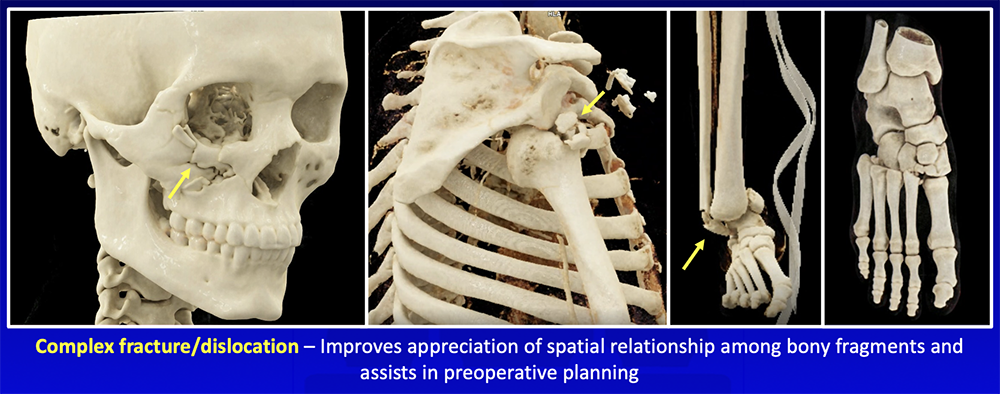 |
Trauma – Vascular Injury 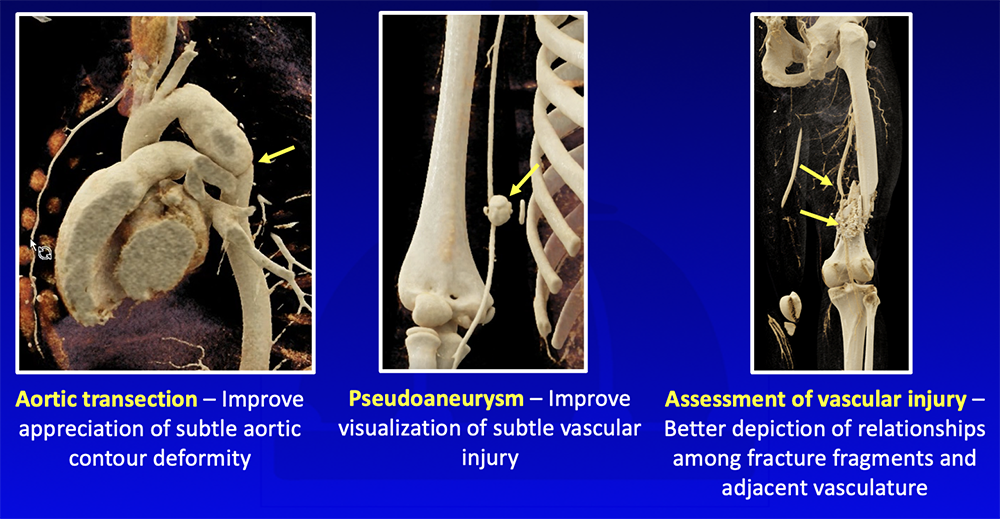 |
Musculoskeletal – Infection/Oncology 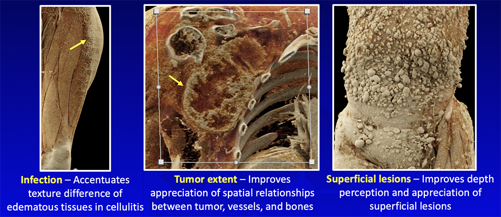 |
Oncologic Applications
|
Oncologic – Lesion Detection and Characterization 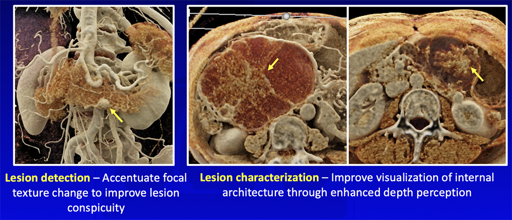 |
Oncologic – Global Disease Burden 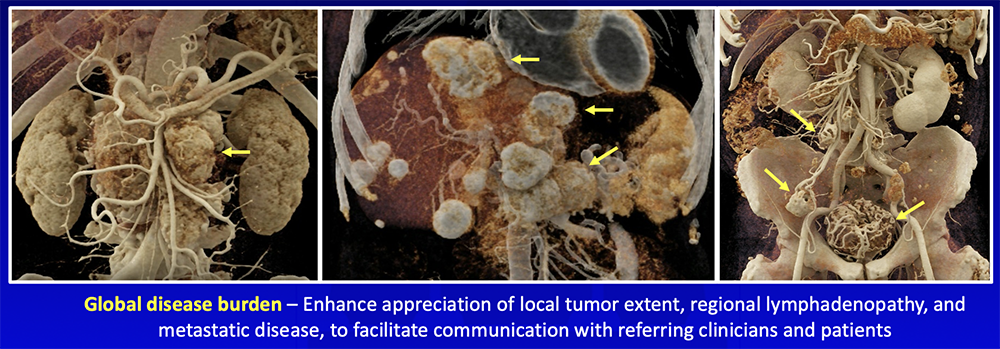 |
Oncologic – Preoperative Planning 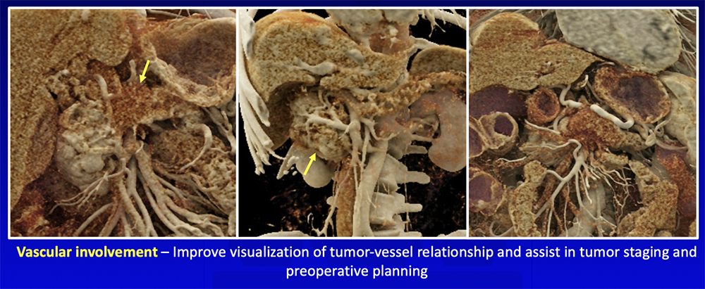 |
Fusion PET/CT Data Rowe SP et al. Abdom Radiol (NY). 2022 [Online ahead of print] 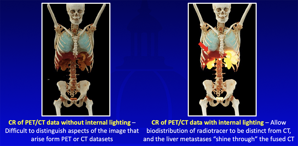 |
How We Do It?
|
How We Do It? The user selects the most appropriate preset (left panel). He or she highlights different tissues and pathologies of interest by adjusting the window, level, and imaging plane. Process took < 1 min. 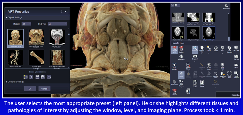 |
How We Do It?
 |
Reimbursement
|
Cinematic Rendering – Value Added
|
Cinematic Rendering – Future Directions Integration with augmented reality
|
Cinematic Rendering with Augmented Reality Rowe SP et al. J Comput Assist Tomogr. 2022 [Online ahead of print]  |
Conclusion
|
References
|