google ads

Taming the Broncho: An Imaging Guide to Acute Bronchial Disorders 

Taming the Broncho: An Imaging Guide to Acute Bronchial Disorders 

Elliot K. Fishman M.D.
Johns Hopkins Hospital


Teaching Points

  • Various bronchial diseases can cause acute symptoms and physiologic abnormalities
  • Apply a systematic approach to analyzing and characterizing bronchial disorders in the emergent setting
  • Disorders can be categorized as follows:
    • Endobronchial lesions
    • Bronchiectasis
    • Bronchial stenosis/wall thickening
    • Bronchomalacia


Differential Dx: Endobronchial Lesions

Benign
  • Aspiration pneumonia
  • Foreign body aspiration
  • Broncholith
  • Pulmonary hamartoma
  • Mucous Plug
  • Infection (tuberculosis, aspergillosis)
  • Lipoma
  • Neurogenic tumor
  • Fibroepithelial polyp
Malignant
  • Carcinoid
  • Adenoid cystic carcinoma
  • Mucoepidermoid carcinoma
  • Metastasis
  • Squamous cell carcinoma
  • Small cell carcinoma
  • Adenocarcinoma
  • Tracheobronchial papillomatosis
  • Lymphoma
José, Cárdenas-García & Lee Chang, Alfredo & Virginia, Chung & Chang, Shim & Factor, Stephen & Amit, Tibb. Bronchial leiomyoma, a case report and review of literature. Respiratory Medicine Case Reports. 2014 May 10;12:59-62.


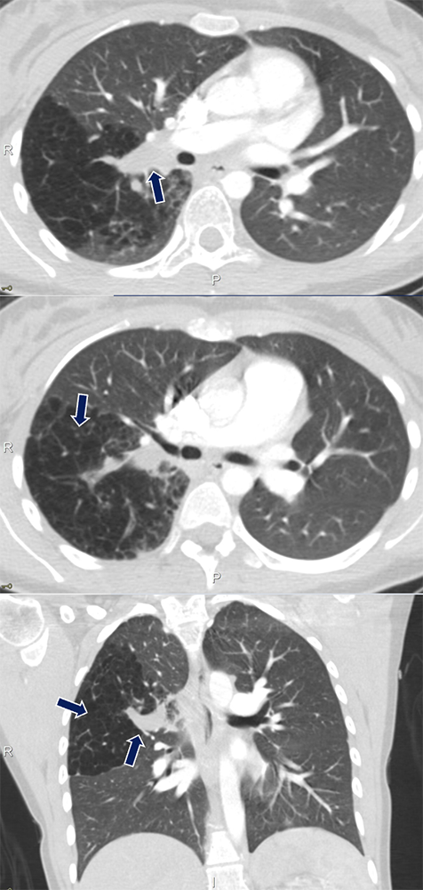
Aspiration Pneumonia

38 yo F with history of respiratory failure status post tracheostomy presenting with tachycardia and dyspnea. Large amount of debris is seen in the bronchus intermedius (arrows) with distal consolidations in the right middle and lower lobes.
  • Wide range of clinical symptoms from asymptomatic to life-threatening with the most common severe complication being from infection due to mixed anaerobic organisms
  • Aspirants typically affect the basal segments, RML and lingula on an upright patient; lower lobe superior segments and upper lobe posterior segments on a supine patients
  • Can have a wide variety of radiologic manifestations depending on the characteristics and volume of the aspirated material and the chronicity of the aspiration
Franquet T, Giménez A, Rosón N, Torrubia S, Sabaté JM, Pérez C. Aspiration diseases: findings, pitfalls, and differential diagnosis. Radiographics. 2000 May-Jun;20(3):673-85.

Aspiration Pneumonia


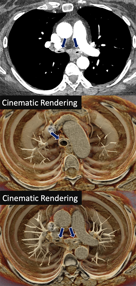
Foreign Body

58 yo F with recent history of pneumonia presenting with sudden onset of dyspnea. An ovoid endobronchial object is seen within the right lower lobe bronchus (arrows). Subsequent bronchoscopy was performed with extraction of an aspirated nut. 
  • Acute obstruction of the airway by an object of variable attenuation depending on its composition
  • No enhancement with contrast; no extraluminal extension
  • Air-trapping, atelectasis, or consolidation may be seen distal to occluded airway
  • Treatment is bronchoscopic extraction
  • Imaging should never delay intervention in life threatening aspirations
Tseng HJ, Hanna TN, Shuaib W, Aized M, Khosa F, Linnau KF. Imaging Foreign Bodies: Ingested, Aspirated, and Inserted. Ann Emerg Med. 2015 Dec;66(6):570-582.e5.

Foreign Body


Foreign Body

Top:76 yo M with aspirated food particle in the right lower lobe bronchus (arrows), retrieved by bronchoscopy
Bottom:9 yo M with cough and chest pain after aspirating a coil spring from a pen. A metallic foreign body (arrows) is lodged within the left mainstem bronchus with partial collapse of the left lung.

Foreign Body


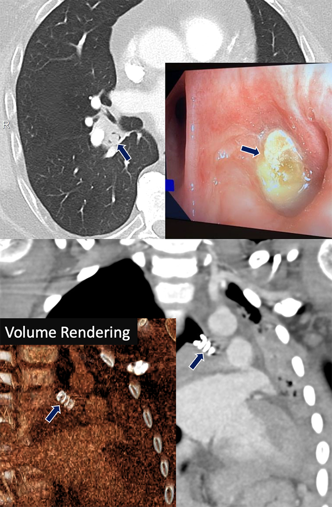
Mucous Plug

Left:72 yo F with large mucous plugging of the entire left-sided airways (arrows) with associated complete left lung collapse. Right:64 yo M with metastatic urothelial cell carcinoma. A small mucous plug was found within a subsegmental bronchus
  • Characteristically appears as a tubular opacity filling the airway
  • Large amount of mucous plugging can cause significant lung collapse
  • Small mucous plugs can be mis-characterized as lung nodules
  • Most commonly associated with asthma, allergic bronchopulmonary aspergillosis, and plastic bronchitis
  • Management usually involves mucolytics, hydration, and humidification but include bronchoscopic intervention
Panchabhai TS, Mukhopadhyay S, Sehgal S, Bandyopadhyay D, Erzurum SC, Mehta AC. Plugs of the Air Passages: A Clinicopathologic Review. Chest. 2016 Nov;150(5):1141-1157.

Mucous Plug


Broncholith

78 yo F with history of lung cancer presenting with left-sided chest pain. A calcified density compatible with broncholith (arrows) is demonstrated in a distal bronchus with proximal mucous plugging and bronchiectasis. Note the ipsilateral calcified nodes.
  • A broncholith is calcified material within bronchial lumen. Can be due to erosion of a calcified lymph node, calcification of previously aspirated material, or from direct aspiration of osseus matter
  • Most commonly presents with cough, hemoptysis, or post-obstructive pneumonia
  • Characteristically appears as a calcified, endobronchial mass with associated obstructive findings such as atelectasis or bronchiectasis
Seo JB, Song KS, Lee JS, Goo JM, Kim HY, Song JW, Lee IS, Lim TH. Broncholithiasis: review of the causes with radiologic-pathologic correlation. Radiographics. 2002 Oct;22 Spec No:S199-213.

Broncholith


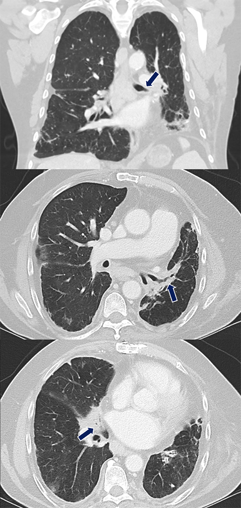
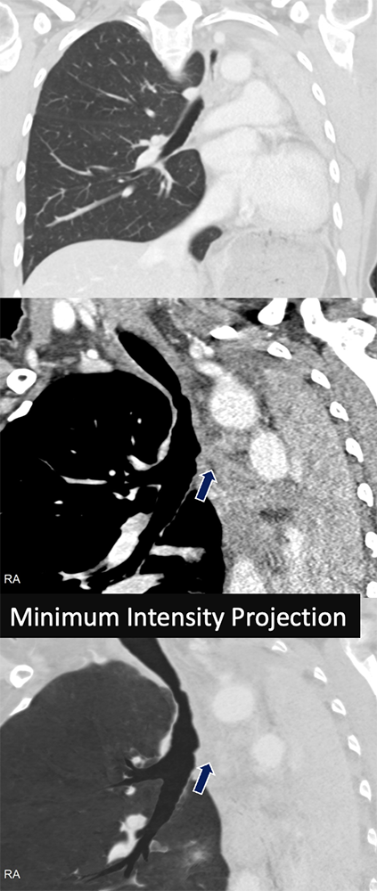
Pulmonary Hamartoma

Top 2: 74 yo F with acute shortness of breath and weakness. A fat-containing lesion (arrows) occludes the left upper lobe bronchus causing complete lobar collapse.
Bottom: Companion case of endobronchial hamartoma in the right lower lobe.
  • Pulmonary hamartomas are benign tumors frequently composed of fat and cartilage 
  • 1.4-20% are endobronchial while the majority are parenchymal
  • Associated with recurrent pneumonia or hemoptysis due to airway compression/obstruction
  • Hamartomas contain variable amount of fat and calcification (popcorn type calcification is classic)
Cosío BG, Villena V, Echave-Sustaeta J, de Miguel E, Alfaro J, Hernandez L, Sotelo T. Endobronchial hamartoma. Chest. 2002 Jul;122(1):202-5.

Pulmonary Hamartoma


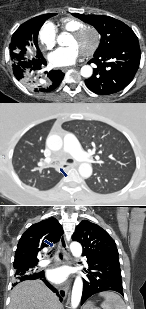
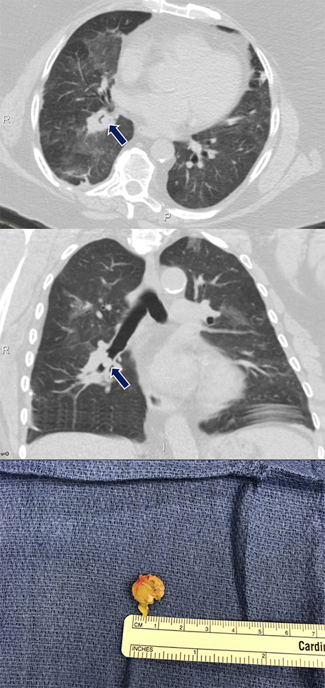
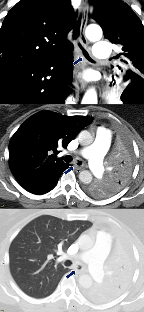
Pulmonary Hamartoma

65 yo F with history of pneumonia causing pain and dyspnea, improved with antibiotics. A small nodular soft tissue lesion (arrows)is seen within the lobar bronchi (left and center). An endobronchial polypoid lesion was found on bronchoscopy (right) at the anterior wall entrance of the right lower lobe bronchus and subsequently resected at its stalk.

Pulmonary Hamartoma


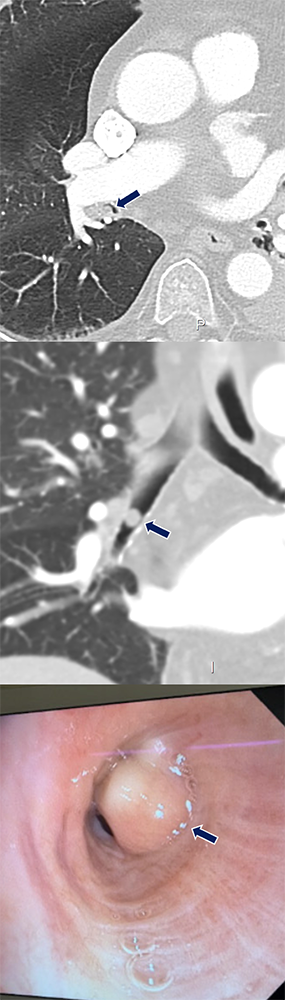
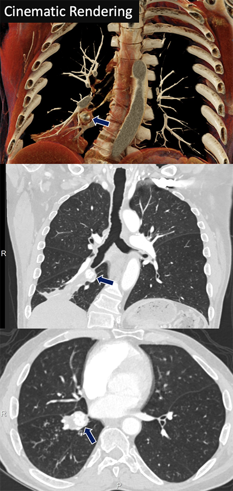
Carcinoid

67 yo M with history of COPD presenting with right lower lobe pneumonia. A coarsely calcified mass (arrows) expands and occludes the right lower lobe bronchus with post-obstructive atelectasis.
  • Carcinoid is a type of neuroendocrine tumors that can arise from bronchial epithelium, often with an endobronchial component; generally considered to be a low-grade (typical carcinoid) or intermediate-grade (atypical carcinoid) malignancy
  • Most commonly present in the pulmonary or GI system but are known to affect all organ systems
  • Carcinoid syndrome (flushing, hypotension, wheezing) only occurs once the malignancy metastasizes to the liver which allows secreted hormones to escape hepatic metabolism
Baxi AJ, Chintapalli K, Katkar A, Restrepo CS, Betancourt SL, Sunnapwar A. Multimodality Imaging Findings in Carcinoid Tumors: A Head-to-Toe Spectrum. Radiographics. 2017 Mar-Apr;37(2):516-536.

Carcinoid


Adenoid Cystic Carcinoma

59 yo F with history of COPD who presented with chest pain and dyspnea. An endobronchial lesion with small calcifications (arrows) is seen in the bronchus intermedius along with collapse of the right lower and middle lobes.
  • Symptoms are related to airway obstruction such as dyspnea, cough, and wheezing.
  • Most commonly arises in the lower trachea but can be found in the bronchi and upper trachea
  • Although often a low-grade malignancy, can invade local tissues such as the thyroid gland
Kwak SH, Lee KS, Chung MJ, Jeong YJ, Kim GY, Kwon OJ. Adenoid cystic carcinoma of the airways: helical CT and histopathologic correlation. AJR Am J Roentgenol. 2004 Aug;183(2):277-81.

Adenoid Cystic Carcinoma


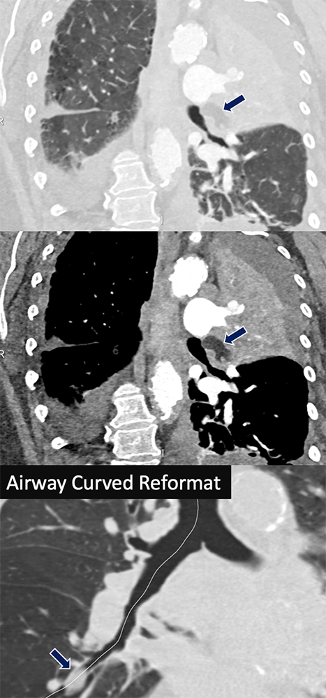
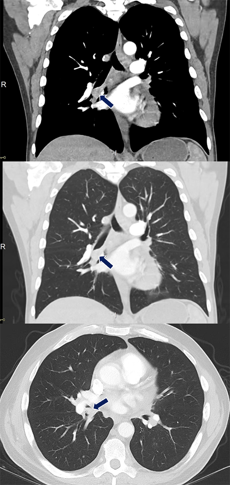
Mucoepidermoid Carcinoma

52 yo F with no PMH who presented with cough, fevers, chills, generalized aches. An endobronchial pedunculated tumor (arrows) occupies the right mainstem bronchus and partially obstructs the lumens of the right upper lobe and bronchus intermedius.
  • Mucoepidermoid carcinoma is a rare, slow-growing malignancy that arises from salivary glands within the tracheobronchial tree
  • Tumor develops within the endoluminal airway space with potential post-obstructive pneumonia or atelectasis
  • The mass typically appears homogenous with mild contrast enhancement and can have associated calcifications
Kim TS, Lee KS, Han J, Im JG, Seo JB, Kim JS, Kim HY, Han SW. Mucoepidermoid carcinoma of the tracheobronchial tree: radiographic and CT findings in 12 patients. Radiology. 1999 Sep;212(3):643-8.

Mucoepidermoid Carcinoma


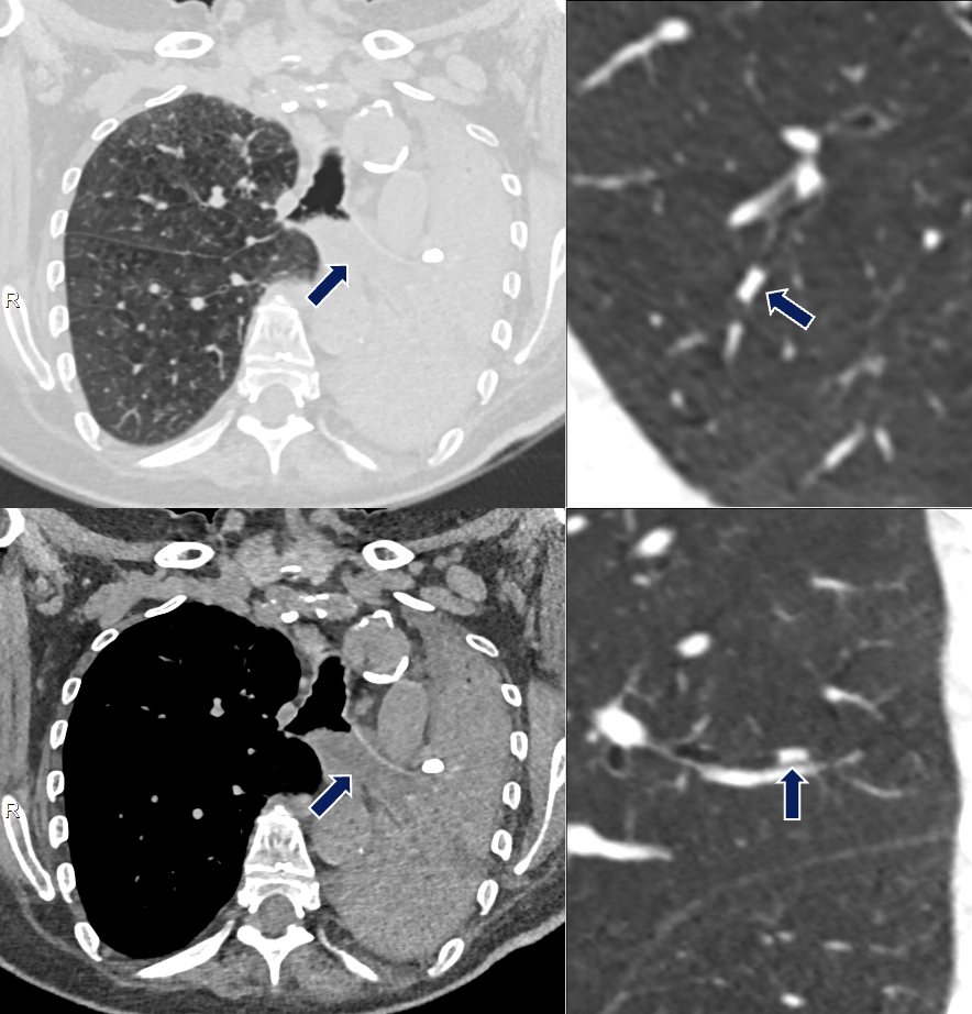
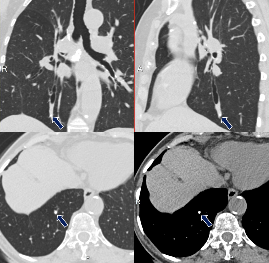
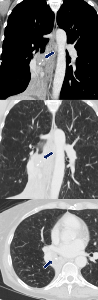
Metastasis from Renal Cell Carcinoma

64 yo M with history of metastatic clear cell carcinoma status post nephrectomy on immunotherapy and chemotherapy. A soft tissue nodule (arrows) developed in the right lower lobe airway later confirmed to be a metastasis.
  • Most common malignancies to metastasize to the bronchus are colorectal, breast, and kidney and can be difficult to distinguish from primary endobronchial malignancy if the primary tumor has not been identified.
  • Often presents in advanced metastatic disease with cough, chest pain, and hemoptysis.
  • Compared with primary endobronchial tumors, metastasis to the bronchus portends a much poorer prognosis
Breta M, Arava S, Madan K, Singh A, Jain D, Guleria R. Endobronchial metastasis from extrathoracic malignancies: A clinicopathological study of 11 cases. Lung India. 2019 May-Jun;36(3):212-215.

Metastasis from Renal Cell Carcinoma


Differential Dx: Bronchial Stenosis/Wall Thickening

  • Bronchial stenosis
  • Bronchial asthma
  • Relapsing polychondritis
  • Tracheobronchial amyloidosis
  • Sarcoidosis
  • Tuberculosis
  • COPD
  • Granulomatous with polyangiitis
  • Infectious bronchitis
Theriault MM, Eddy K, Borgaonkar JN, Babar JL, Manos D. Diseases Involving the Central Bronchi: Multidetector CT for Detection, Characterization, and Differential Diagnosis. Radiographics. 2018 Jan-Feb;38(1):58-59.


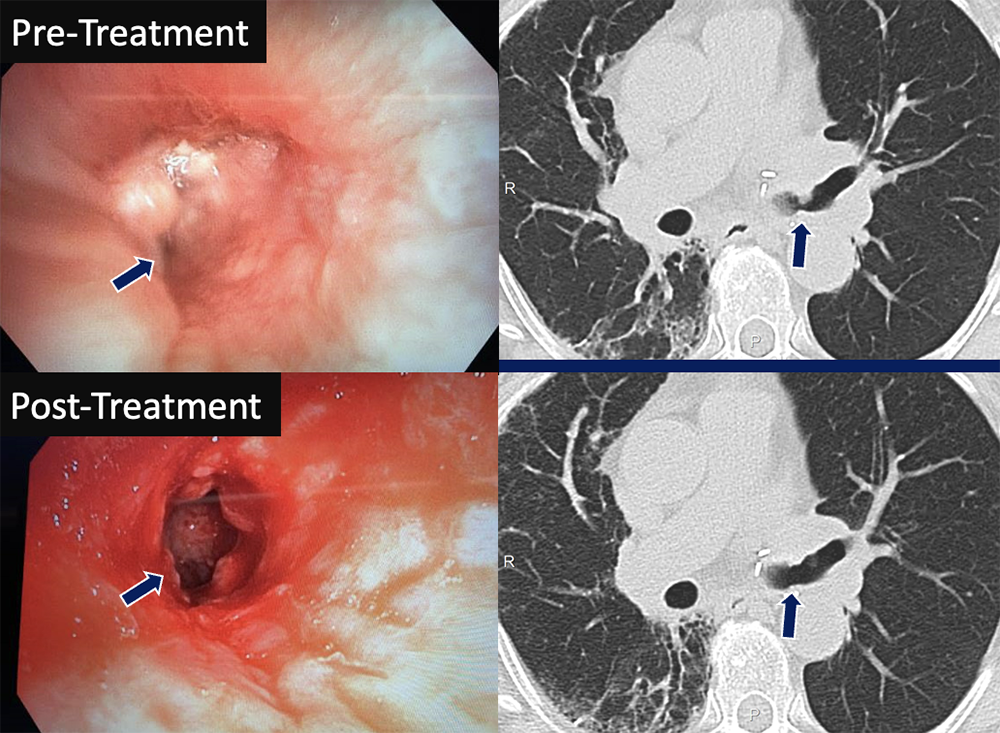
Bronchial Stenosis

74 yo F status post left lung transplant with decline in PFTs. CT and pre-treatment bronchoscopy images (left) showed anastomotic narrowing at the left mainstem bronchus, worse on expiration. A cryoprobe was used to debride exophytic granulation tissue in the airway followed by balloon dilation of the bronchus. The left mainstem bronchus regained patency on post-treatment images (right).
  • Multitude of causes ranging from idiopathic, iatrogenic (long term intubation or tracheostomy), infection, and systemic disease
  • Distribution of the disease, associated findings, and presence of airway calcifications can narrow the differential diagnosis
  • When associated bronchomalacia is present, degree of stenosis will worsen with expiration and therefore dynamic airway CT can be diagnostic
Grenier PA, Beigelman-Aubry C, Brillet PY. Nonneoplastic tracheal and bronchial stenoses. Radiol Clin North Am. 2009 Mar;47(2):243-60.

Bronchial Stenosis


Bronchial Asthma

54 yo M presenting with acute cough, wheezing, and fatigue. Segmental and subsegmental bronchi (arrows), particularly in the lower lobes, are diffusely thickened and narrowed.
  • Bronchial wall thickening is common in severe asthma
  • Indicates smooth muscle hypertrophy, inflammation, and fibrosis
  • The degree of thickening and stenosis correlates with disease severity
Walker C, et al. Computed tomography scans in severe asthma: utility and clinical implications. Curr Opin Pulm Med. 2012;18(1):42-47. 

Bronchial Asthma


Relapsing Polychondritis

35 yo F presenting with hemoptysis and chest pain. The trachea and bilateral mainstem bronchi show diffuse and smooth wall thickening (arrows) with sparing of the posterior membrane and associated luminal narrowing.
  • Rare autoimmune disorder characterized by recurrent inflammation of cartilaginous structures in the body such as the tracheobronchial tree, larynx, joints, and nose
  • Appears as anterior and lateral wall thickening of the airway due to fibrotic changes caused by recurrent inflammation, sparing the posterior membrane
  • Respiratory failure accounts for 50% of deaths due to relapsing polychondritis
Lee KS, Ernst A, Trentham DE, Lunn W, Feller-Kopman DJ, Boiselle PM. Relapsing polychondritis: prevalence of expiratory CT airway abnormalities. Radiology. 2006 Aug;240(2):565-73. Epub 2006 Jun 26.

Relapsing Polychondritis


Tracheobronchial Amyloidosis

62 yo F with history of amyloidosis presenting with dyspnea. Images show marked circumferential soft tissue thickening (arrows) of the walls of the distal trachea extending into the right mainstem bronchus with associated narrowing and partially collapsed left lung.
  • Amyloidosis is a multisystem disorder of abnormal protein deposition. Tracheobronchial manifestations include airway wall thickening and intramural nodules that can be complicated by airway obstruction
  • Airway involvement can be circumferential including the posterior membrane unlike cartilaginous disorders (ie. relapsing polychondritis and tracheobronchopathia osteochondroplastica)
  • Disease of the upper airway presents with typical upper airway symptoms while disease of the lower airways presents with recurrent pneumonia and lobar collapse
Czeyda-Pommersheim, F., Hwang, M., Chen, S. S., Strollo, D., Fuhrman, C., & Bhalla, S. (2015). Amyloidosis: Modern Cross-sectional Imaging. Radiographics : a review publication of the Radiological Society of North America, Inc, 35(5), 1381–1392.

Tracheobronchial Amyloidosis


Sarcoidosis

60 yo M with history of multisystem sarcoidosis presents with chronic cough and baseline dyspnea. Multifocal bronchial wall thickening and stenosis (arrows)was noted in the left lower lobe, lingula, and right middle lobe bronchus with right middle lobe collapse.
  • Multisystem, autoimmune disorder with formation of noncaseous granulomas. Pulmonary involvement is seen in 90% of patients and accounts for a majority of the morbidity and mortality of the disease
  • Common presenting symptoms include cough, dyspnea, and fatigue although many patients with sarcoidosis are asymptomatic. 
  • More than half of patients experience minimal consequences from the disease but around one fifth of patients can develop pulmonary fibrosis.
Criado, E., Sánchez, M., Ramírez, J., Arguis, P., de Caralt, T. M., Perea, R. J., & Xaubet, A. (2010). Pulmonary sarcoidosis: typical and atypical manifestations at high-resolution CT with pathologic correlation. Radiographics : a review publication of the Radiological Society of North America, Inc, 30(6), 1567–1586.

Sarcoidosis


Tuberculosis

43 yo F with history of treated tuberculosis. The left lung was completely collapsed with fibrotic/atretic appearance of the left mainstem bronchus (arrows). Note the associated volume loss and mediastinal shift.
  • Initial infection with Mycobacterium tuberculosis is referred to as primary tuberculosis and can appear radiographically as mediastinal and hilar lymphadenopathy, consolidation, pleural effusion, or nodules. 
  • Following resolution of the primary infection, tuberculosis can appear as upper lung consolidations and cavitations.
  • Both primary and post-primary disease can affect the bronchial wall and manifests as bronchial wall involvement and bronchial stenosis due to lymphadenitis.
Nachiappan, A. C., Rahbar, K., Shi, X., Guy, E. S., Mortani Barbosa, E. J., Jr, Shroff, G. S., Ocazionez, D., Schlesinger, A. E., Katz, S. I., & Hammer, M. M. (2017). Pulmonary Tuberculosis: Role of Radiology in Diagnosis and Management. Radiographics : a review publication of the Radiological Society of North America, Inc, 37(1), 52–72.

Tuberculosis


Differential Dx: Bronchiectasis

Focal
  • Bronchial atresia
  • Extrinsic compression
  • Sarcoidosis
  • Post-radiation fibrosis
  • Repeated aspiration
Diffuse
  • Nontuberculous mycobacterial infection
  • Pseudomonas infection
  • Cystic fibrosis
  • Allergic bronchopulmonary aspergillosis
  • Graft versus Host disease
  • Primary ciliary dyskinesia
  • Tracheobronchomegaly
  • Williams-Campbell Syndrome
  • Hypogammaglobulinemia
Cantin L, Bankier AA, Eisenberg RL. Bronchiectasis. AJR Am J Roentgenol. 2009 Sep;193(3):W158-71.


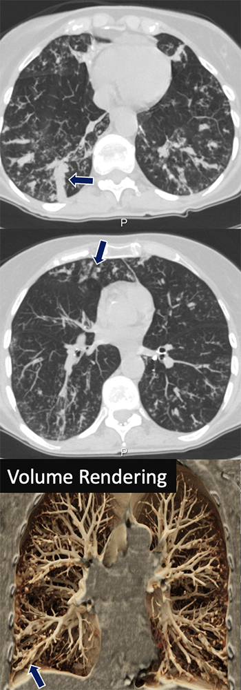
Bronchial Atresia

20 yo F presents with acute chest pain. A low density tubular lesion occludes the right upper lobe segmental bronchus with marked distal air-trapping (arrows).
  • Congenital abnormality of the bronchial tree causing focal region of stenosis thought to be secondary to intrauterine trauma or ischemic injury
  • CT imaging often shows overinflated alveoli in lung parenchyma adjacent to the affected bronchus with a mucocele just distal to the stenotic region 
  • Clinical presentation can vary from asymptomatic to dyspnea and recurrent pulmonary infections. 
Gipson, M. G., Cummings, K. W., & Hurth, K. M. (2009). Bronchial atresia. Radiographics : a review publication of the Radiological Society of North America, Inc, 29(5), 1531–1535.

Bronchial Atresia


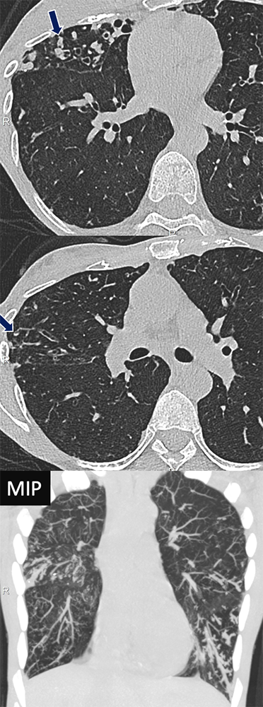
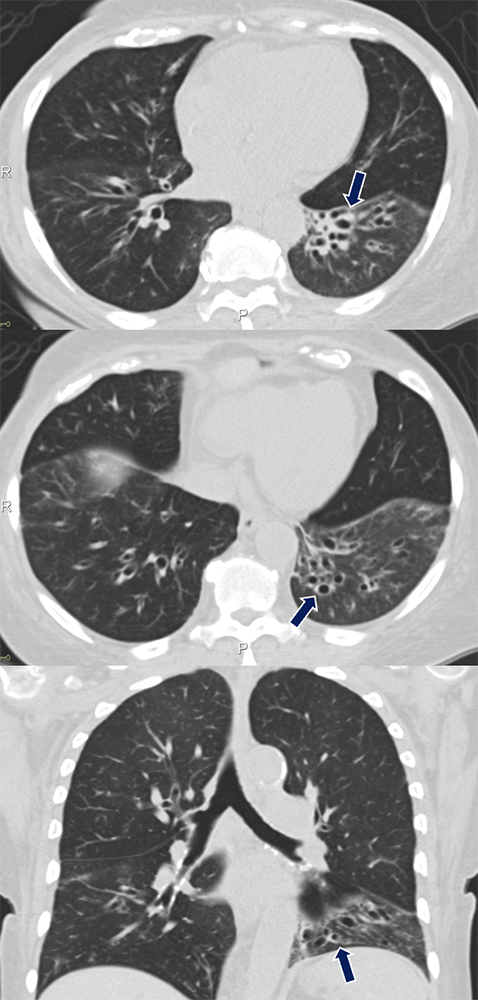
Nontuberculous Mycobacterial Infection

67 yo F with idiopathic bronchiectasis presenting with worsening productive cough, fevers, and chills. Mucoceles seen as dilated tubular structures that fills the airway spaces with mucus (arrows), worse at the lower lobes. Abnormal bronchial wall thickening and centrilobular tree-in-bud nodularity representing involvement of the bronchioles and alveolar spaces.
  • Typically occurs in older women (Lady Windermere Syndrome) and presents with chronic cough
  • CT imaging usually shows peripheral bronchiectasis, bronchial wall thickening, tree-in-bud nodules, and larger random nodules that may cavitate
  • Usually spares the upper lungs; classic location is in the right middle lobe and lingula
Erasmus JJ, McAdams HP, Farrell MA, Patz EF Jr. Pulmonary nontuberculous mycobacterial infection: radiologic manifestations. Radiographics. 1999 Nov-Dec;19(6):1487-505.

Nontuberculous Mycobacterial Infection


Pseudomonas Infection

63 yo F with recurrent pseudomonas infections who presents with worsening productive cough. Extensive bronchiectasis, bronchial wall thickening, tubular mucous plugging, and centrilobular tree-in-bud nodularity was found (arrows), better seen on MIP.
  • Common cause of hospital-acquired pneumonia, particularly amongst patients on ventilators
  • Nosocomial pseudomonal pneumonia carries a 60-80% mortality rate making rapid diagnosis and management especially important
  • CT imaging classically shows bilateral airspace opacities with ground glass opacities, bronchiectasis, mucous plugging, and bronchial wall thickening
Okada, F., Ono, A., Ando, Y., Nakayama, T., Ishii, R., Sato, H., Kira, A., Tokimatsu, I., Kadota, J., & Mori, H. (2012). Thin-section CT findings in Pseudomonas aeruginosa pulmonary infection. The British journal of radiology, 85(1020), 1533–1538.

Pseudomonas Infection


Cystic Fibrosis

25 yo F with cystic fibrosis presenting with chest pain. CT showing cystic and varicose atelectasis and areas of bronchial wall thickening and mucous plugging (arrows).
  • Cystic fibrosis is the most common genetic disorder of white children and has historically had a poor prognosis. However, the recent advent of CFTR modulators has improved the expected lifespan and quality of life of patients
  • Detection and progression of the disease is best characterized with CT and often shows upper lung predominant bronchiectasis, wall thickening, mucous plugging, and nodularity
  • Patients with cystic fibrosis often have repeated bouts of pneumonia with drug-resistant organisms such as Pseudomonas, S. Aureus
Ernst, C. W., Basten, I. A., Ilsen, B., Buls, N., Van Gompel, G., De Wachter, E., Nieboer, K. H., Verhelle, F., Malfroot, A., Coomans, D., De Maeseneer, M., & de Mey, J. (2014). Pulmonary disease in cystic fibrosis: assessment with chest CT at chest radiography dose levels. Radiology, 273(2), 597–605.
Lopes-Pacheco M. CFTR Modulators: The Changing Face of Cystic Fibrosis in the Era of Precision Medicine. Front Pharmacol. 2020 Feb 21;10:1662.

Cystic Fibrosis


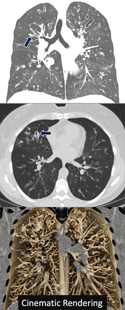
Allergic Bronchopulmonary Aspergillosis

54 yo F with presenting with productive cough, prior history of dyspnea and intermittent chest tightness. CT demonstrated multiple tubular mucous plugging seen as Y-shaped finger-in-glove opacities involving the central bronchi (arrows).
  • Typically occurs in patients with long standing asthma or cystic fibrosis
  • Presents with wheezing and pneumonia-like symptoms of productive cough and fever 
  • Imaging classically shows the "finger-in-glove" sign of dilated tubular branching opacities which demonstrates mucus plugging
  • Attenuation of the mucus plug will exceed that of skeletal muscle on noncontrast CT
Thompson BH, Stanford W, Galvin JR, Kurihara Y. Varied radiologic appearances of pulmonary aspergillosis. Radiographics. 1995 Nov;15(6):1273-84. 
Franquet T, Müller NL, Giménez A, Guembe P, de La Torre J, Bagué S. Spectrum of pulmonary aspergillosis: histologic, clinical, and radiologic findings. Radiographics. 2001 Jul-Aug;21(4):825-37.  

Allergic Bronchopulmonary Aspergillosis


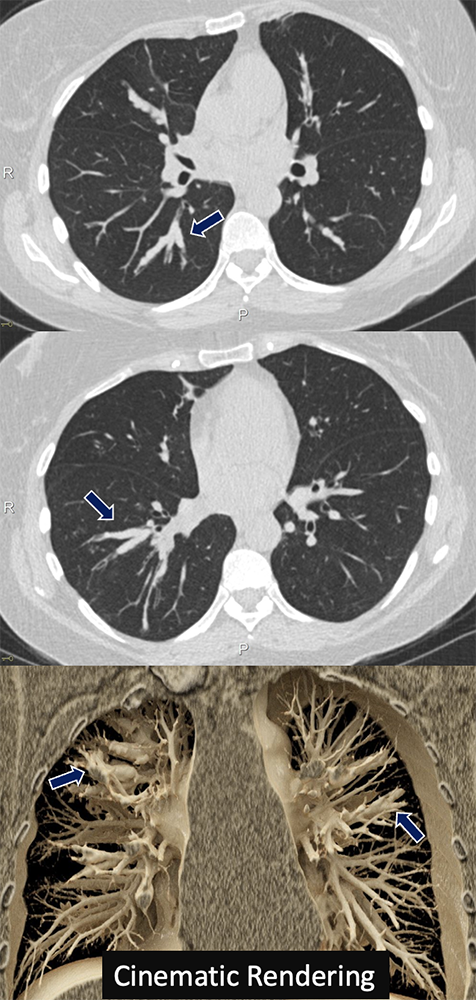
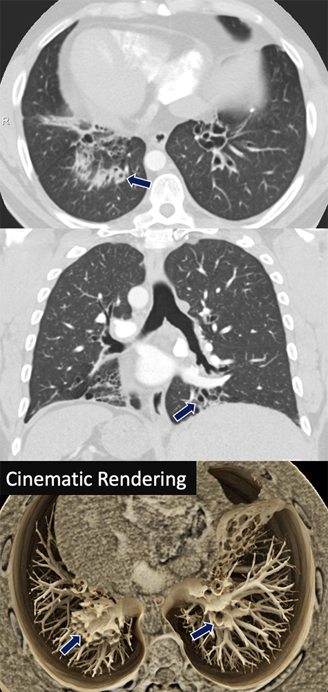
Graft Versus Host Disease

66 yo F with history of AML s/p bone marrow transplant who presented with hypoxia and cough. CT scan showed significant bronchiectasis and bronchial wall thickening most severe in the left lower lobe (arrows).
  • GVHD is a serious complication from hematopoietic stem cell transplantation and can occur acutely after transplantation or with more chronic progression.
  • Chronic GVHD within the pulmonary system is referred to as bronchiolitis obliterans and manifests as bronchial inflammation and fibrosis leading to small airway narrowing.
  • CT is the ideal modality for assessing for bronchiolitis obliterans and shows a mosaic pattern of ventilation and perfusion. CT can also show lower lung predominant bronchiectasis, bronchial wall thickening, and air trapping.
Peña, E., Souza, C. A., Escuissato, D. L., Gomes, M. M., Allan, D., Tay, J., & Dennie, C. J. (2014). Noninfectious pulmonary complications after hematopoietic stem cell transplantation: practical approach to imaging diagnosis. Radiographics : a review publication of the Radiological Society of North America, Inc, 34(3), 663–683.

Graft Versus Host Disease


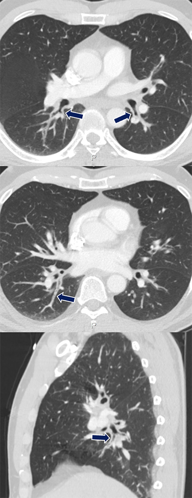
Primary Ciliary Dyskinesia

34 yo M with history of primary ciliary dyskinesia (Kartagener's syndrome) who presents with productive cough and fever. Marked bronchial wall thickening and bronchiectasis are seen in the lower lungs (arrows). Note the associated cardiac situs inversus.
  • Primary ciliary dyskinesia is caused by abnormal ciliary function leading to impaired mucous clearance within the airway. Kartagener's Syndrome describes a subset of patients with a triad of impaired ciliary clearance, situs inversus, and sinusitis
  • Clinical symptoms are similar to that of cystic fibrosis with chronic respiratory infection being the primary cause of morbidity and mortality. 
  • CT imaging classically shows severe bronchiectasis and bronchial wall thickening.
Kennedy, M. P., Noone, P. G., Leigh, M. W., Zariwala, M. A., Minnix, S. L., Knowles, M. R., & Molina, P. L. (2007). High-resolution CT of patients with primary ciliary dyskinesia. AJR. American journal of roentgenology, 188(5), 1232–1238.

Primary Ciliary Dyskinesia


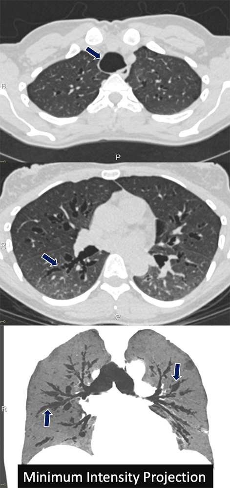
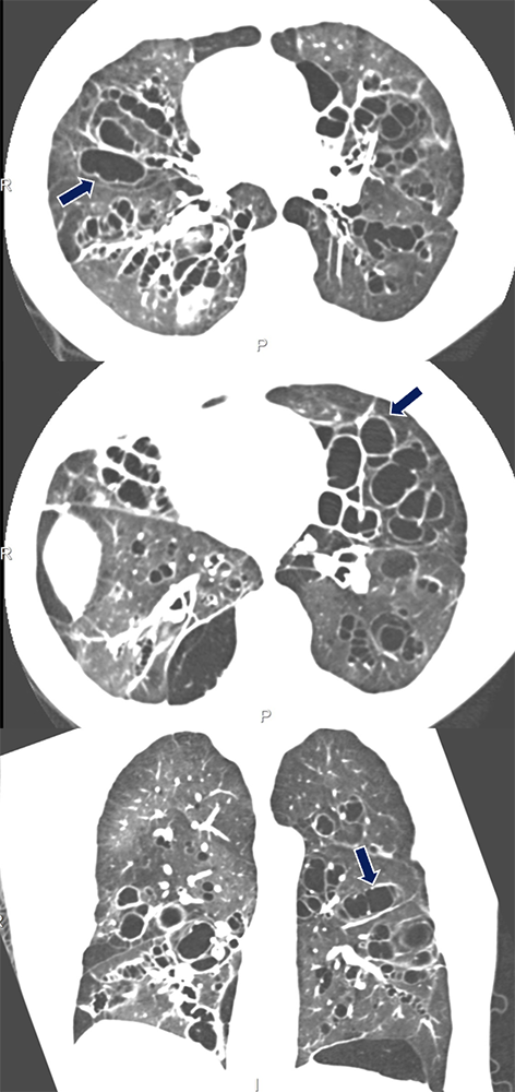
Tracheobronchomegaly

36 yo F with history of recurrent pneumonia who presented with confusion and shortness of breath. Imaging shows diffuse dilatation of the trachea and proximal bronchi with segments of varicose bronchiectasis (arrows).
  • Tracheobronchomegaly, also referred to as Mounier-Kuhn Syndrome, is a disease of trachea and bronchial dilation due to atrophy of elastic tissue. It is poorly understood but thought to be a sporadic genetic disorder.
  • Patients have a wide range of clinical presentations from asymptomatic to severe respiratory failure. Due to poor mucociliary clearance recurrent lower respiratory tract infections are common.
  • Dilation of the trachea and central bronchi is often accompanied by tracheal and bronchial diverticula
Celik, B., Bilgin, S., & Yuksel, C. (2011). Mounier-Kuhn syndrome: a rare cause of bronchial dilation. Texas Heart Institute journal, 38(2), 194–196. 

Tracheobronchomegaly


Williams-Campbell Syndrome

40 yo M previously diagnosed with asthma presenting with cough, wheezing, and shortness of breath. Branching dilated airways with thickened walls characteristic of bronchiectasis (arrows), involving the mid-order bronchi. 
  • Rare disease caused by defective cartilage in the 4th-6th generation bronchi leading to central cystic bronchiectasis
  • Congenital form that classically presents in children but can sometimes present in adulthood
  • Bronchi will dilate on inspiration and collapse on expiration (bronchomalacia)
Noriega Aldave AP, William Saliski D. The Clinical Manifestations, Diagnosis and Management of Williams-Campbell Syndrome. N Am J Med Sci. 2014 Sep;6(9):429-32.  Ghosh S. Williams-Campbell Syndrome. Radiology. 2022 Feb;302(2):274.

Williams-Campbell Syndrome


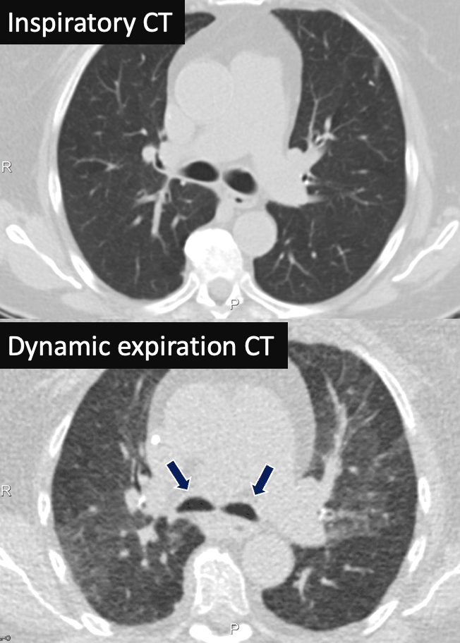
Bronchomalacia

82 yo F presenting with persistent cough. Inspiratory CT show patent and normal-sized bilateral mainstem bronchi. Dynamic CT during expiration show dynamic collapse of the bilateral mainstem bronchi (arrows) with associated air trapping.
  • Bronchomalacia is a part of a group of disorders that cause dynamic airway collapse on expiration due to weakness of the supportive cartilage in the airway.
  • Often accompanies other pulmonary disorders such as asthma or COPD
  • Assessment is best done with dynamic bronchoscopy or dynamic CT imaging
Aslam, A., De Luis Cardenas, J., Morrison, R. J., Lagisetty, K. H., Litmanovich, D., Sella, E. C., Lee, E., & Agarwal, P. P. (2022). Tracheobronchomalacia and Excessive Dynamic Airway Collapse: Current Concepts and Future Directions. Radiographics : a review publication of the Radiological Society of North America, Inc, 42(4), 1012–1027.

Bronchomalacia


Bronchomalacia

76 yo M with cough and shortness of breath, intermittent for several years. Inspiratory chest CT demonstrated patent bilateral mainstem bronchi. On bronchoscopy, the airways were inspected and notable for dynamic airway collapse. Expiratory images show 80% dynamic airway obstruction.
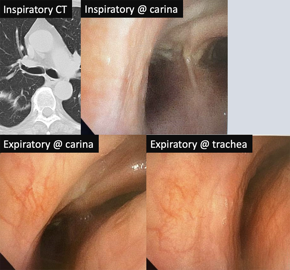
Bronchomalacia


Conclusion

  • Radiologists should be aware of potential pathology that affect the bronchial lumen, size, or wall thickness
  • Bronchial disorders range in presentation from asymptomatic to insidious onset to respiratory distress 
  • Post-processing techniques, dynamic airway CT, and bronchoscopy should be considered in the evaluation and management of affected patients


Privacy Policy

Copyright © 2024 The Johns Hopkins University, The Johns Hopkins Hospital, and The Johns Hopkins Health System Corporation. All rights reserved.