Cinematic Rendering of Chest Anatomy: a Pattern Based Approach
Cinematic Rendering of Chest Anatomy: a Pattern Based Approach Hannah S. Recht, MD Hannah Ahn, MA Elliot K. Fishman, MD The Russel H. Morgan Department of Radiology and Radiological Science The Johns Hopkins Medical Institutions Baltimore, MD, USA |
 |
Tutorial Mode
|
Lungs and Airways |
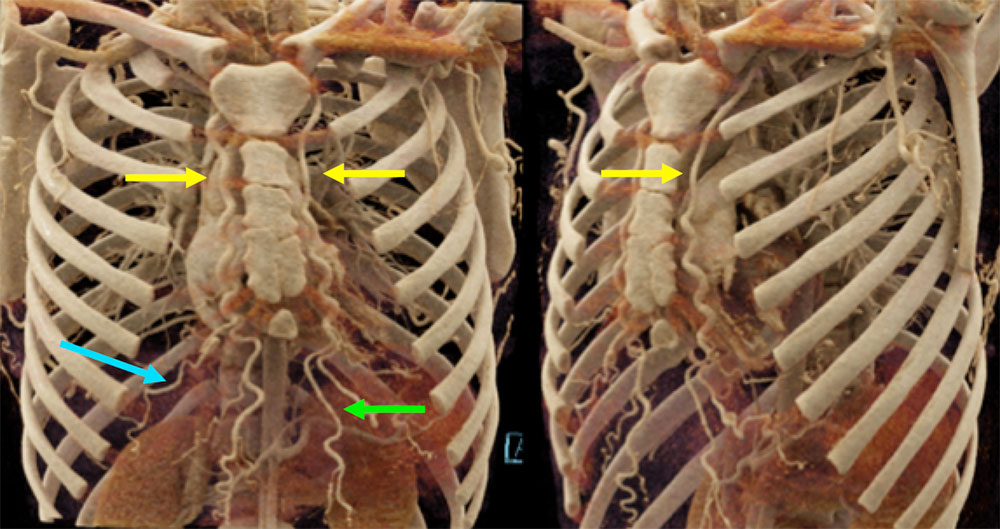
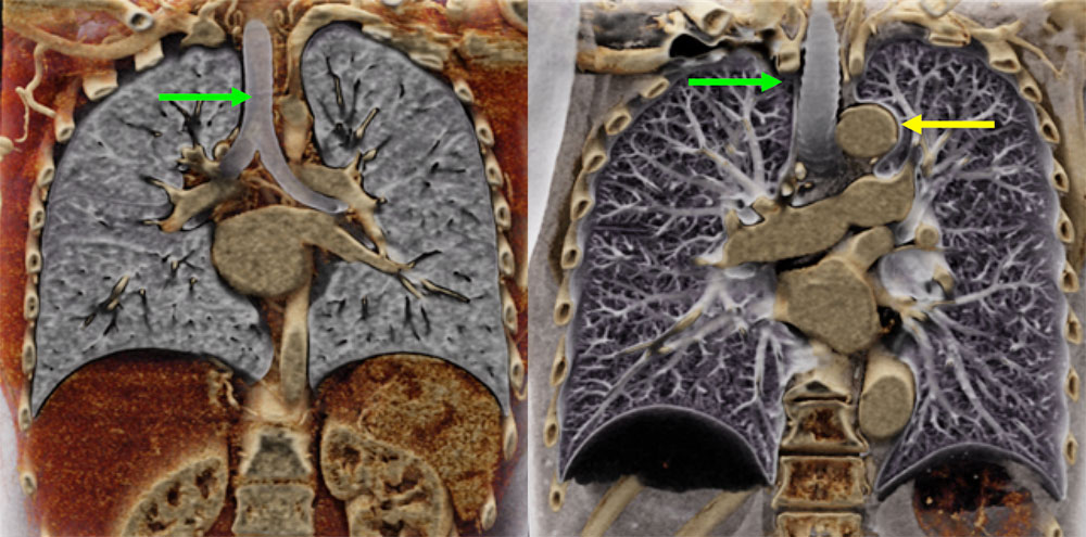 These two coronal CR images show the relationship between the trachea (green arrow), mainstem bronchi, and the adjacent great vessels (yellow arrow). The textural contrasts between different organs is also highlighted, which is pronounced given the significant variation between the air filled lungs and more solid adjacent organs. These two coronal CR images show the relationship between the trachea (green arrow), mainstem bronchi, and the adjacent great vessels (yellow arrow). The textural contrasts between different organs is also highlighted, which is pronounced given the significant variation between the air filled lungs and more solid adjacent organs. |
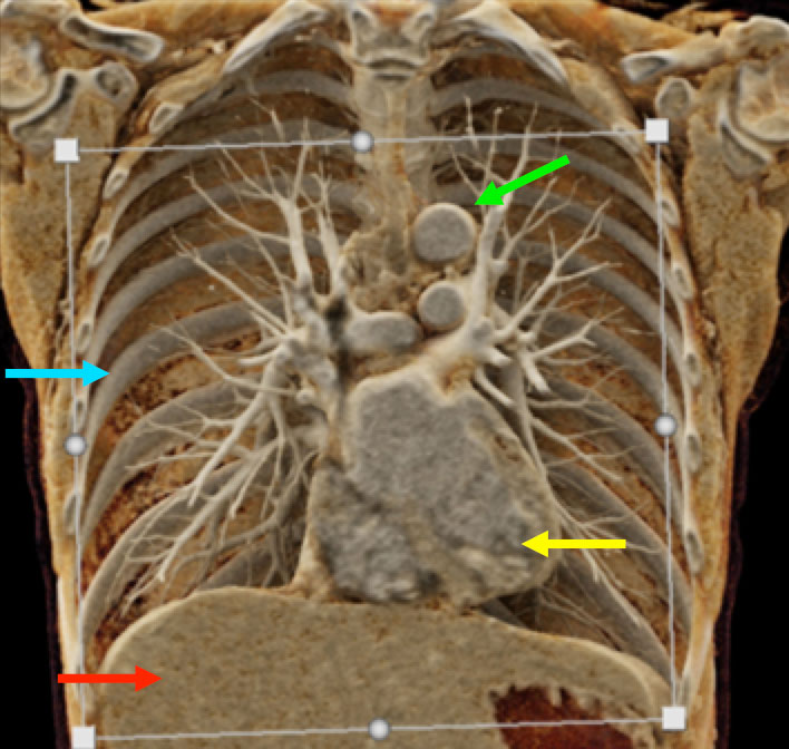
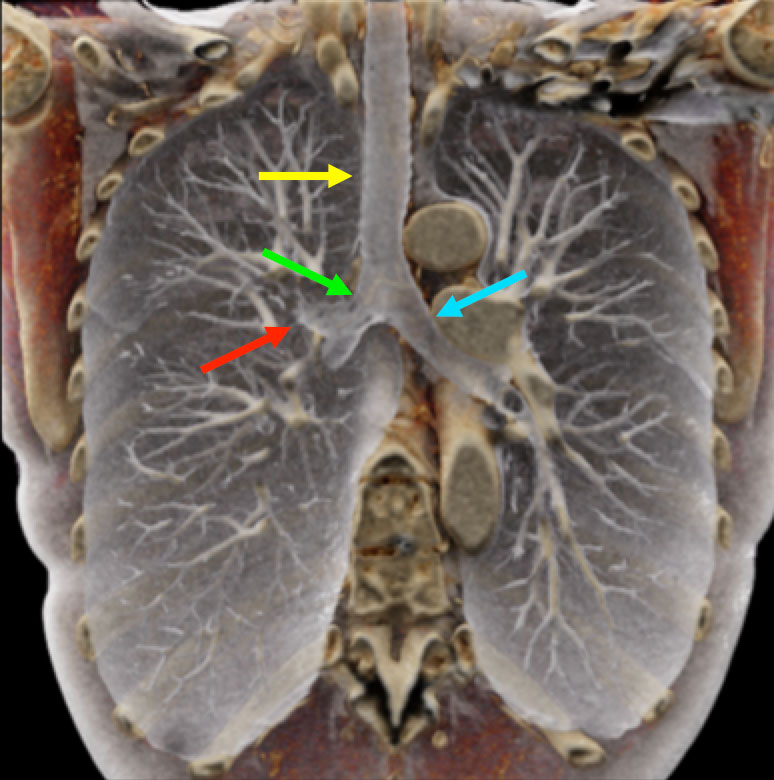 This coronal CR through the lung fields demonstrates the unique photorealistic images that cinematic rendering can provide. The air filled trachea (yellow arrow), right mainstem bronchus (green arrow), left mainstem bronchus (blue arrow), and right upper lobar bronchus (red arrow) are clearly visualized, as are both the right and left lungs. This coronal CR through the lung fields demonstrates the unique photorealistic images that cinematic rendering can provide. The air filled trachea (yellow arrow), right mainstem bronchus (green arrow), left mainstem bronchus (blue arrow), and right upper lobar bronchus (red arrow) are clearly visualized, as are both the right and left lungs. |
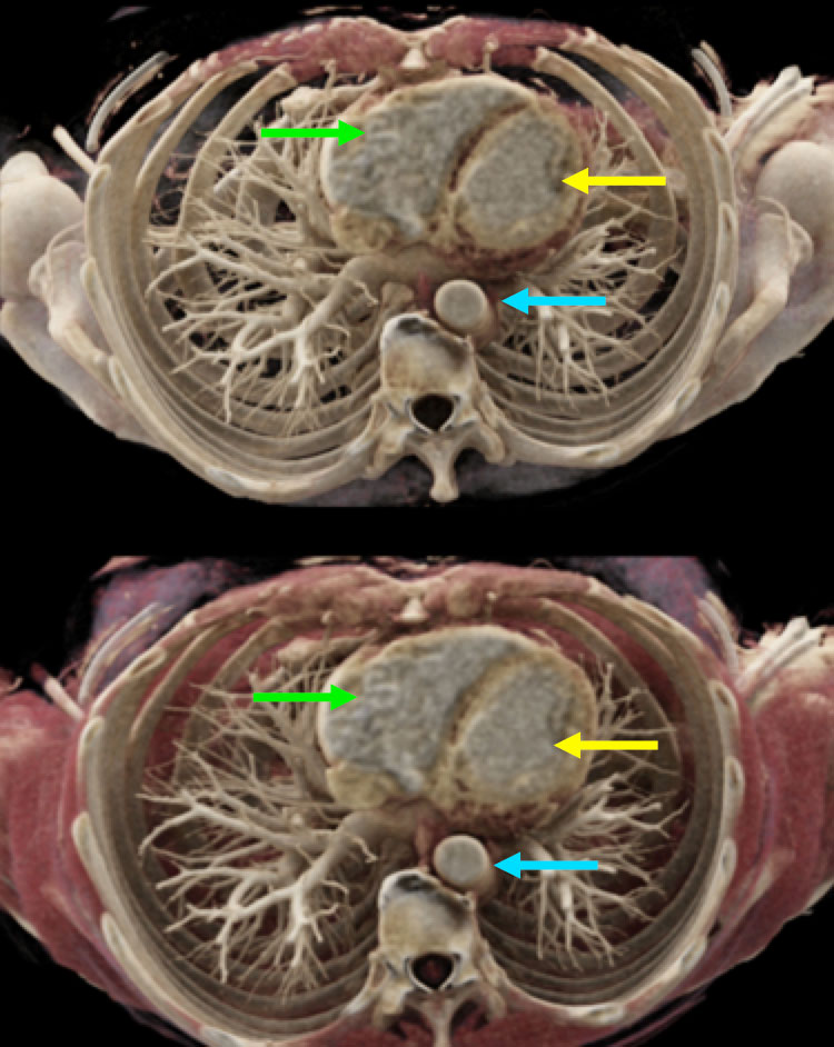
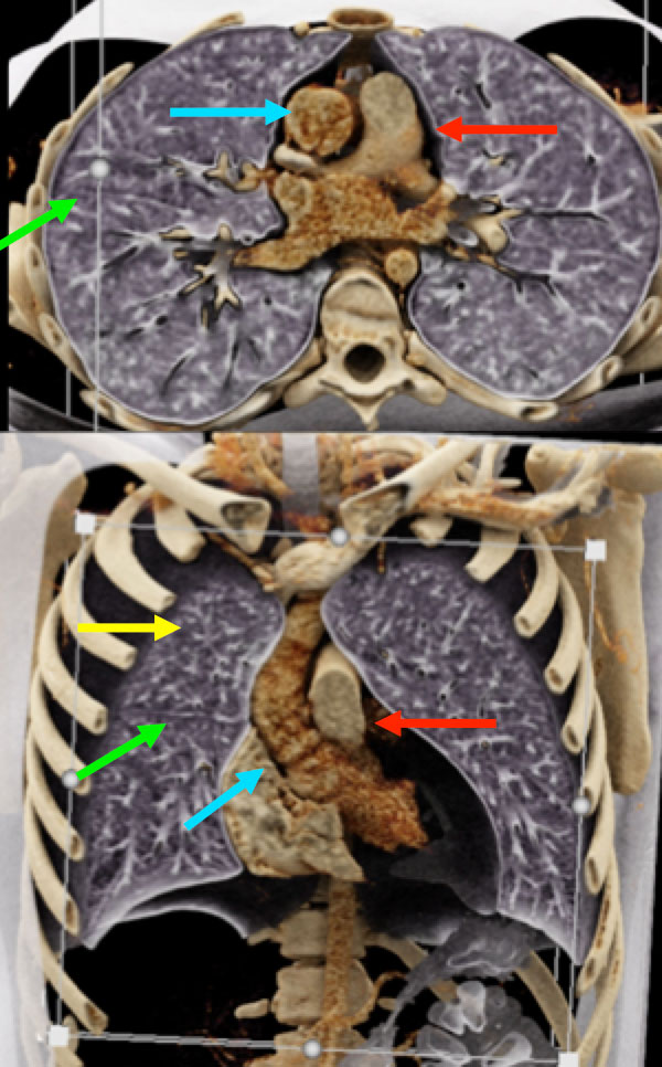 These axial and coronal CR images further highlight the textural differences between the lung fields and mediastinal structures. The images demonstrate enough fine detail that even the fine right minor fissure can be seen. These axial and coronal CR images further highlight the textural differences between the lung fields and mediastinal structures. The images demonstrate enough fine detail that even the fine right minor fissure can be seen. Yellow arrow – right upper lobe Green arrow – right minor fissure Blue arrow – ascending aorta Red arrow – main pulmonary artery |
Vessels |
Vessels |
 These coronal and coronal oblique CR images highlight the course of the paired internal thoracic (internal mammary) arteries (yellow arrow) as they descend inferiorly along the anterior chest wall. The internal thoracic artery has two terminal branches, the musculophrenic artery (blue arrow) and the superior epigastric artery (green arrow). These coronal and coronal oblique CR images highlight the course of the paired internal thoracic (internal mammary) arteries (yellow arrow) as they descend inferiorly along the anterior chest wall. The internal thoracic artery has two terminal branches, the musculophrenic artery (blue arrow) and the superior epigastric artery (green arrow). |
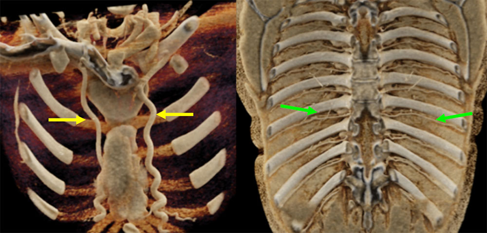 These two coronal CR images highlight the unique capability of CR to provide unique views that cannot be obtained by standard MDCT to highlight specific structures. In the left image, internal thoracic arteries (yellow arrows) are seen from a unique posterior approach. The right CR image isolates the intercostal arteries (green arrows), and demonstrates their subcostal course. These two coronal CR images highlight the unique capability of CR to provide unique views that cannot be obtained by standard MDCT to highlight specific structures. In the left image, internal thoracic arteries (yellow arrows) are seen from a unique posterior approach. The right CR image isolates the intercostal arteries (green arrows), and demonstrates their subcostal course. |
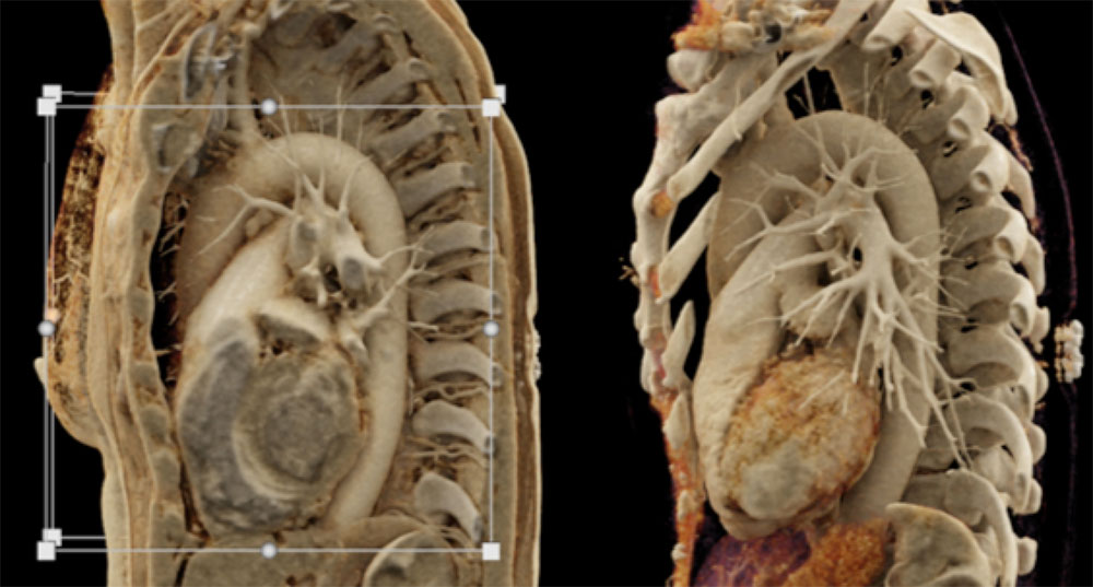 These sagittal oblique CR images demonstrate another distinctive capability of CR imaging. The relative depth of thoracic structures can be visualized, as seen spanning from the anterior to posterior chest wall. Certain structures can also be selected out, as seen in the right CR image, drawing focus to the heart and great vessels. These sagittal oblique CR images demonstrate another distinctive capability of CR imaging. The relative depth of thoracic structures can be visualized, as seen spanning from the anterior to posterior chest wall. Certain structures can also be selected out, as seen in the right CR image, drawing focus to the heart and great vessels. |
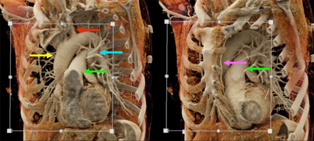 In addition to assessment of depth, these CR images also highlight textural differences between organs. In addition to assessment of depth, these CR images also highlight textural differences between organs. Yellow arrow – ascending thoracic Green arrow – main pulmonary artery Blue arrow – pulmonary veins Red arrow – left subclavian artery Purple arrow – internal thoracic artery |
Cardiac |
 This coronal CR image presents a cross sectional view at the level of the ventricles. Despite being a cross sectional view at one level, CR is able to depict the intrathoracic structures in three dimensions, allowing for intraventricular evaluation, as well as visualization of the adjacent vessels, musculoskeletal structures, and even upper abdomen. This coronal CR image presents a cross sectional view at the level of the ventricles. Despite being a cross sectional view at one level, CR is able to depict the intrathoracic structures in three dimensions, allowing for intraventricular evaluation, as well as visualization of the adjacent vessels, musculoskeletal structures, and even upper abdomen.Yellow arrow – left ventricle Green arrow – aortic arch Blue arrow – posterior rib Red arrow – liver |
 While these axial CR images are presented in a way that is similar to standard MDCT, the CR images provide three dimensional and textural information that standard that standard MDCT cannot. While these axial CR images are presented in a way that is similar to standard MDCT, the CR images provide three dimensional and textural information that standard that standard MDCT cannot. Yellow arrow – left ventricle Green arrow – right ventricle Blue arrow – descending thoracic aorta |
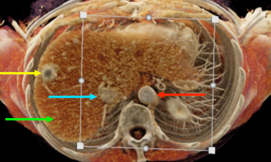 This axial CR image demonstrates the ability of CR to highlight textural contrasts between structures. The liver parenchyma (green arrow) is seen distinct from the IVC (blue arrow) and descending thoracic aorta (red arrow). Incidental note is made of a hepatic metastasis (yellow arrow) in this patient with medullary thyroid cancer. This axial CR image demonstrates the ability of CR to highlight textural contrasts between structures. The liver parenchyma (green arrow) is seen distinct from the IVC (blue arrow) and descending thoracic aorta (red arrow). Incidental note is made of a hepatic metastasis (yellow arrow) in this patient with medullary thyroid cancer. |
Muscles |
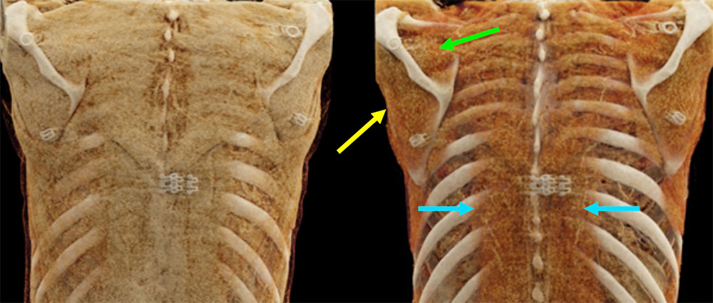
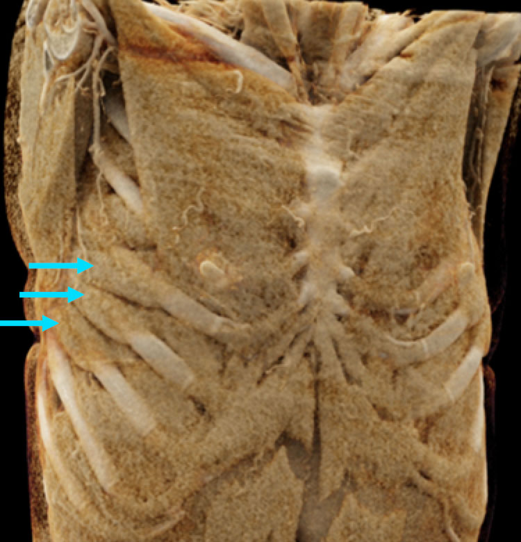 The individual muscle groups are well illustrated by cinematic rendering. The photorealistic images allow visualization of complex muscular origins and insertions. For example, the serratus anterior muscle (blue arrows) is seen attaching to the outer aspect of the upper ribs. Given its size and location, this muscle is not well evaluated on standard MDCT. The individual muscle groups are well illustrated by cinematic rendering. The photorealistic images allow visualization of complex muscular origins and insertions. For example, the serratus anterior muscle (blue arrows) is seen attaching to the outer aspect of the upper ribs. Given its size and location, this muscle is not well evaluated on standard MDCT. |
 These coronal CR images demonstrate the ability of CR to isolate and highlight deep structures, improving visualization of complex anatomy. The right image highlights the paraspinal muscles (blue arrows) in the absence of the larger muscle groups of the back. The supraspinatus (green arrow) and infraspinatus muscles (yellow arrow) are also well seen at their scapular attachments. These coronal CR images demonstrate the ability of CR to isolate and highlight deep structures, improving visualization of complex anatomy. The right image highlights the paraspinal muscles (blue arrows) in the absence of the larger muscle groups of the back. The supraspinatus (green arrow) and infraspinatus muscles (yellow arrow) are also well seen at their scapular attachments. |
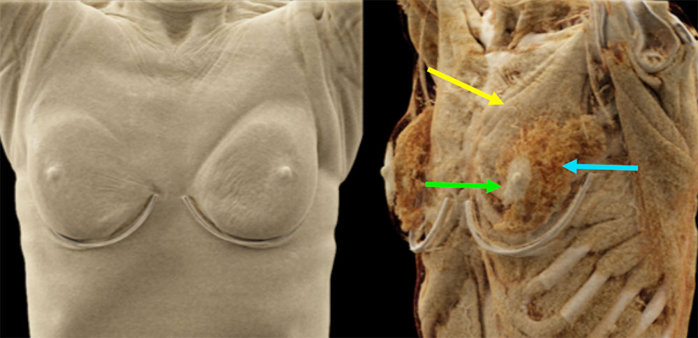 CR can provide three dimensional surface rendering of the skin surface, as well as provide improved anatomic detail of subcutaneous tissues and their relationship to the adjacent organs. The areola (green arrow) is seen distinct from the breast tissue (blue arrow). Both are seen superficial to the pectoralis major muscle (yellow arrow), demonstrating how CR can provide information on relative depth. CR can provide three dimensional surface rendering of the skin surface, as well as provide improved anatomic detail of subcutaneous tissues and their relationship to the adjacent organs. The areola (green arrow) is seen distinct from the breast tissue (blue arrow). Both are seen superficial to the pectoralis major muscle (yellow arrow), demonstrating how CR can provide information on relative depth. |
Bones |
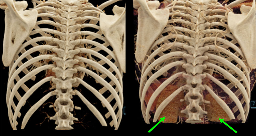 The photorealistic images of CR do an excellent job demonstrating osseous anatomy. The three dimensional images also provide excellent visualization of the relationships between the osseous structures and the adjacent organs. On the right coronal CR image, the kidneys (green arrows) are seen lying ventral to the ribs and spine, showing how trauma to the ribs can lead to renal injury. The photorealistic images of CR do an excellent job demonstrating osseous anatomy. The three dimensional images also provide excellent visualization of the relationships between the osseous structures and the adjacent organs. On the right coronal CR image, the kidneys (green arrows) are seen lying ventral to the ribs and spine, showing how trauma to the ribs can lead to renal injury. |
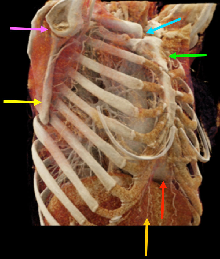 Textural differences and relative depth of anatomic structures as they relate to the axial and appendicular skeleton are highlighted by this oblique coronal CR image. Textural differences and relative depth of anatomic structures as they relate to the axial and appendicular skeleton are highlighted by this oblique coronal CR image. Yellow arrow – scapula Green arrow – manubrium of the sternum Blue arrow – sternoclavicular joint Purple arrow – glenohumeral joint Red arrow – heart Orange arrow – liver |
References
|