- 3
- 3
- ,
- 6
- 8
- 4
To Quiz Yourself: Select OFF by clicking the button to hide the diagnosis & additional resources under the case.
Quick Browser: Select ON by clicking the button to hide the additional resources for faster case review.
CASE NUMBER
5,509


Diagnosis
Cirrhosis with Large Varices and Portal Vein Thrombosis
Note
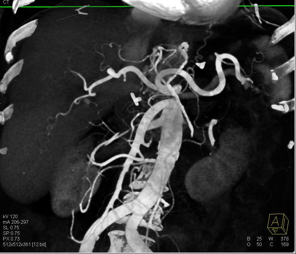
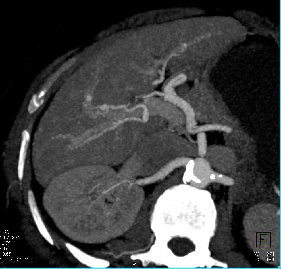
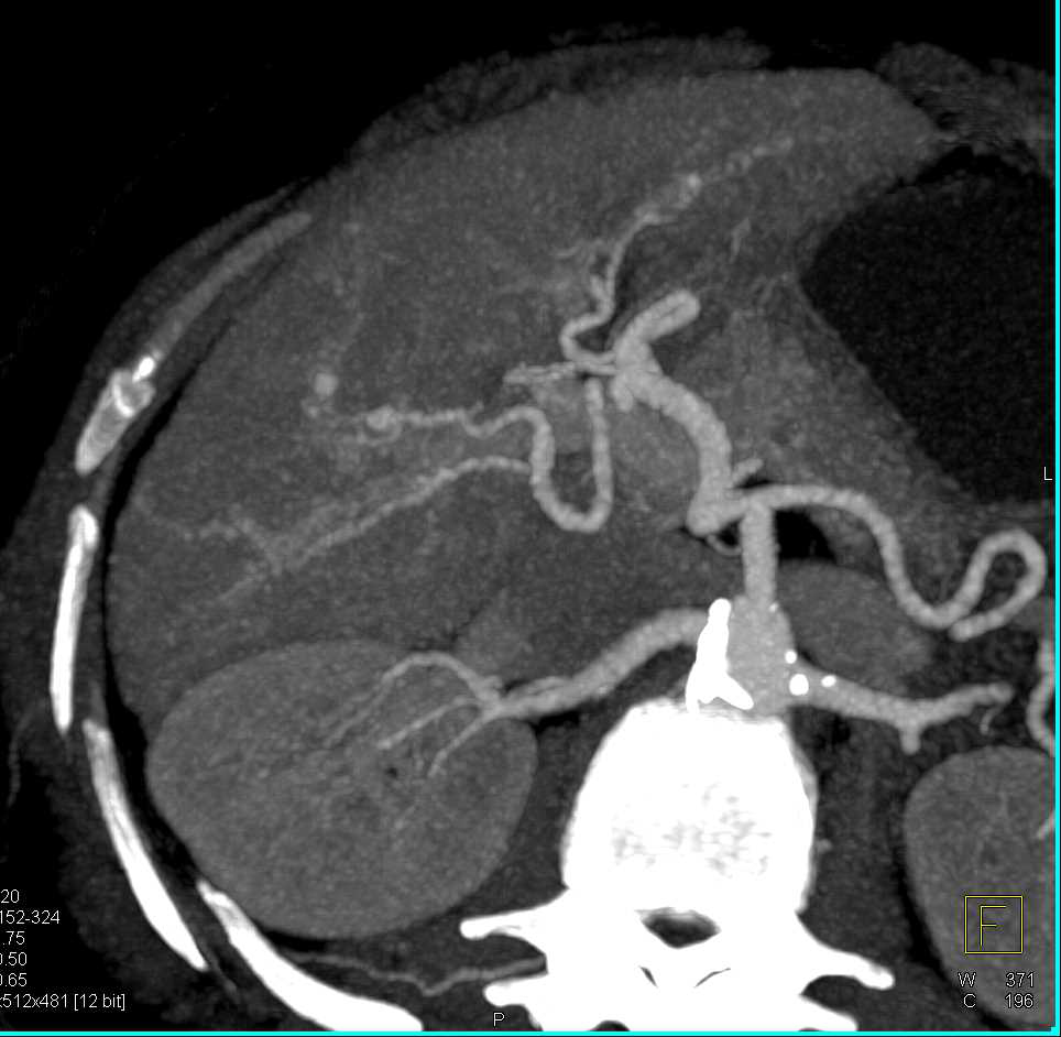
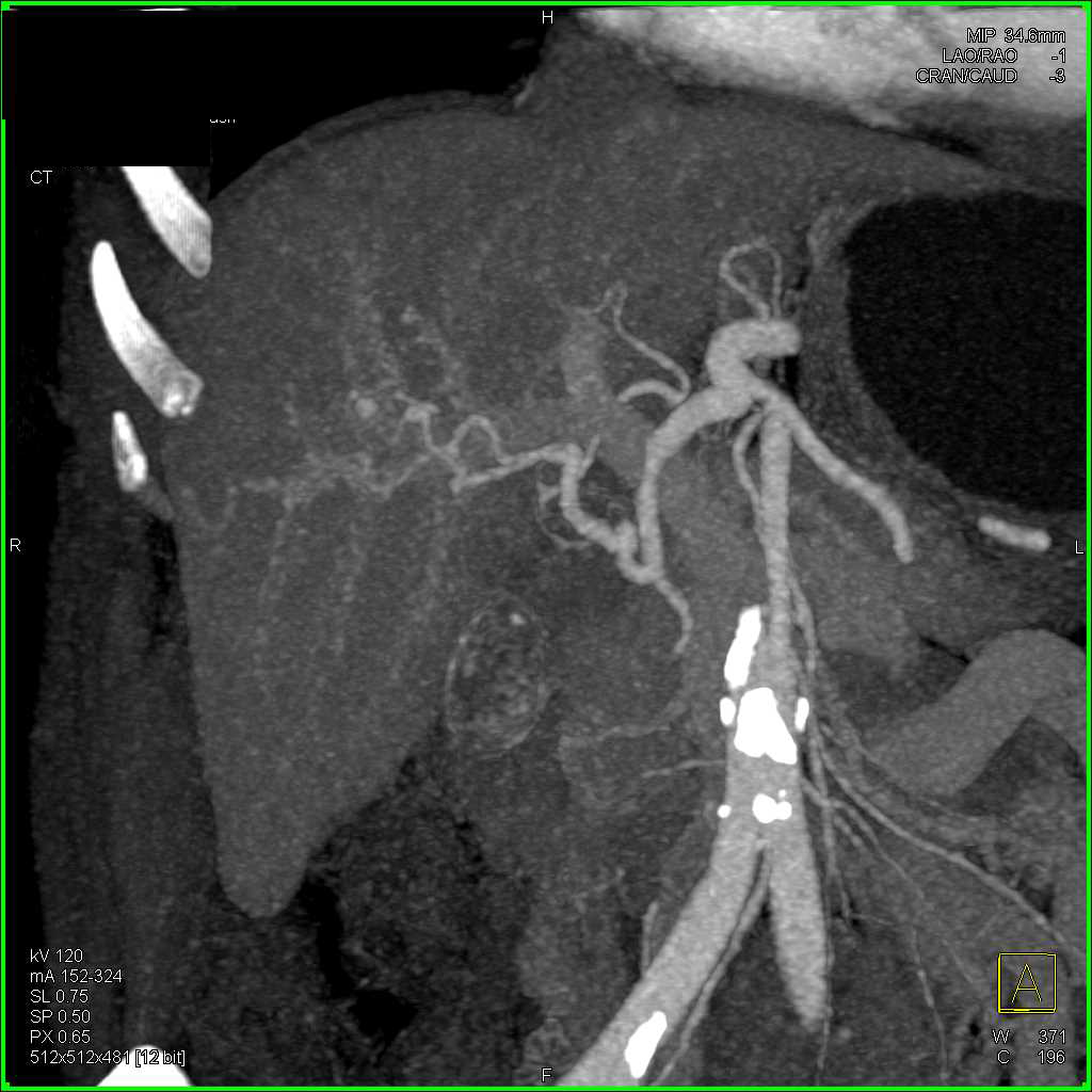
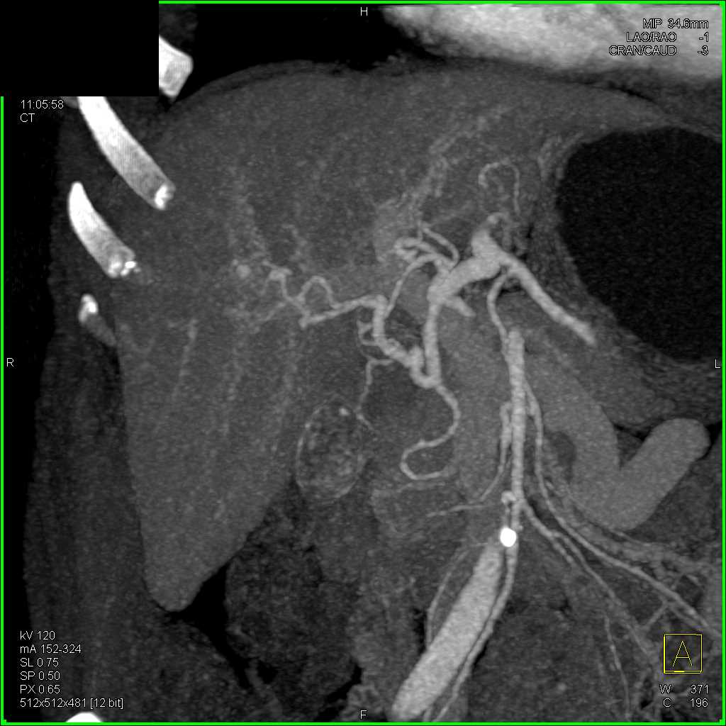
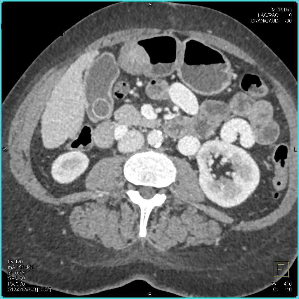
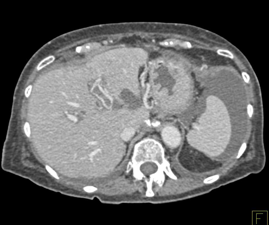
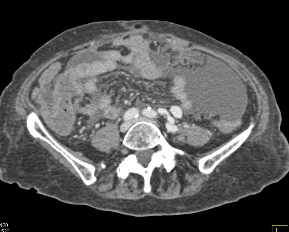
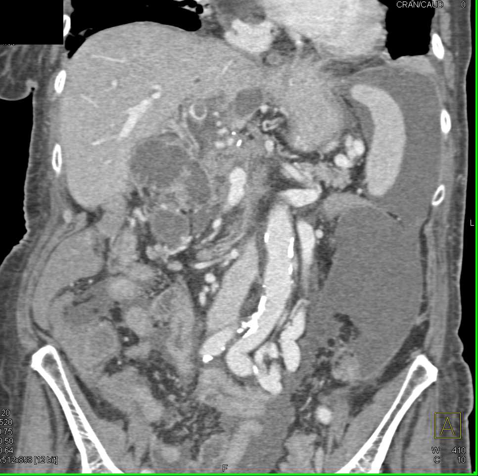
Cirrhosis with large varices and portal vein thrombosis. I think all of us like to look at the liver. The livers challenging. Often you see lesions that are classic: FNH, hepatic adenoma, hepatoma things like that, perhaps. But you can get AV shunting in the liver, small aneurysms in patients with cirrhosis. You can see here there are several small microaneurysms off the hepatic artery. The liver texture is irregular.
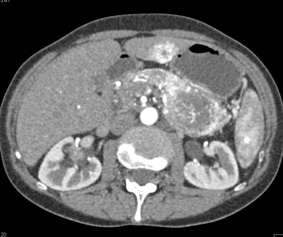
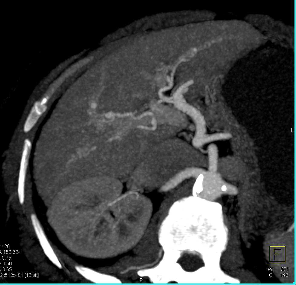
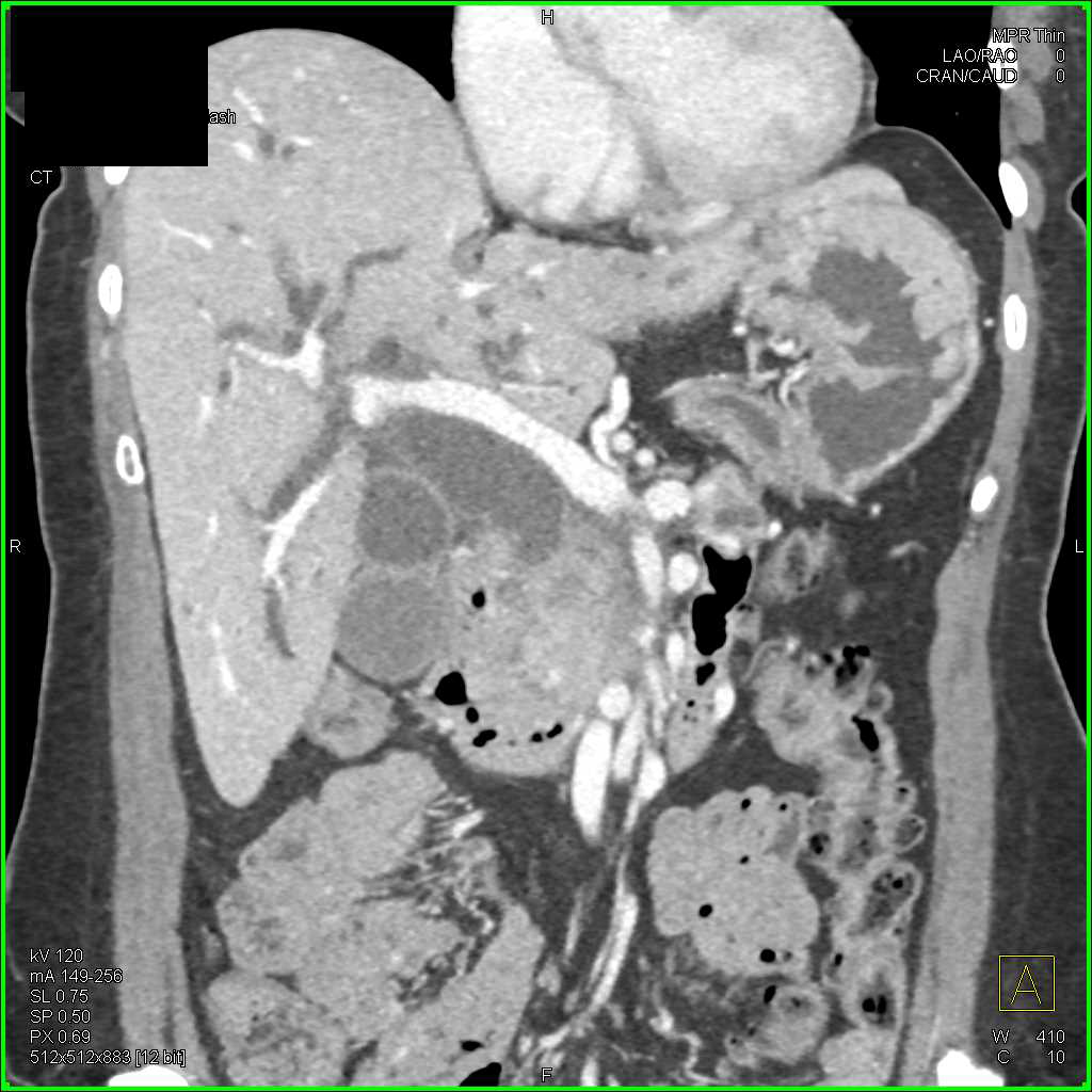
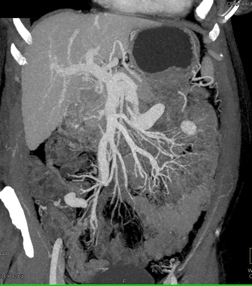
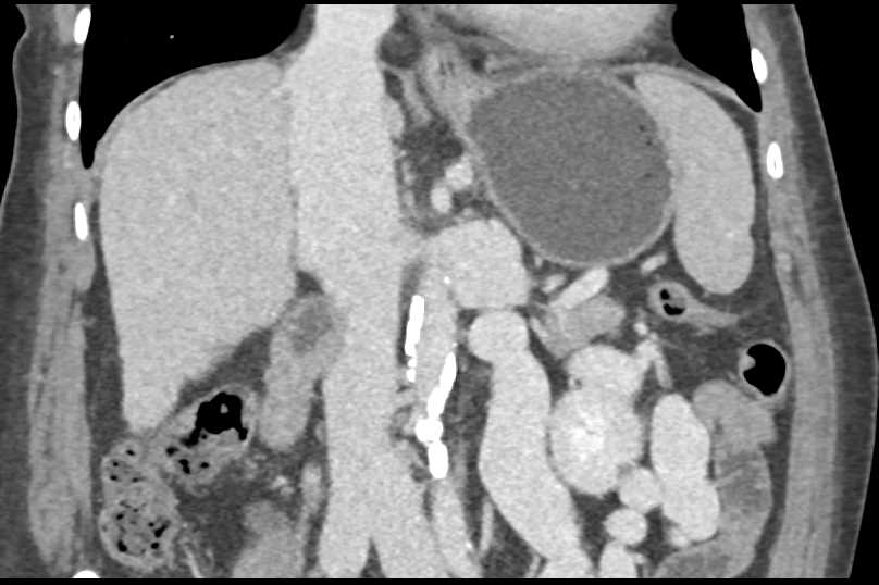
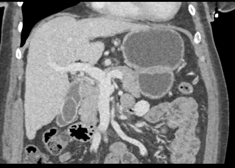
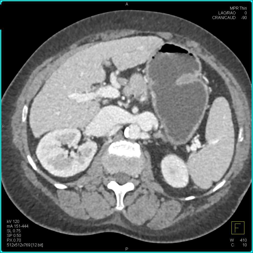
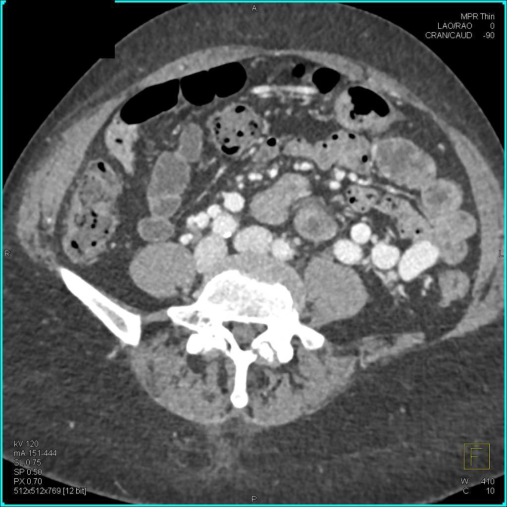
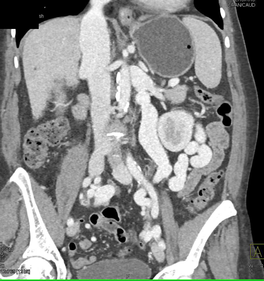
Now obviously youre not going to see portal vein thrombosis or the venous changes well on a MIP image from the arterial phase, so when you look at the venous phase imaging, you see the splenorenal shunting. You see the portal vein looking patent. You see the SMV, you see the gallstone. But as you go higher up, now you can see the thrombus in the portal vein. You can see ascites present, fluid around the spleen.
So I think one of the things that we always need to look at is look at the portal vein. Portal vein thrombosis can be due to hypercoagulability states. It can be due to tumor extension like pancreatic cancer. It also can be seen with hepatoma and cholangiocarcinoma, often by direct tumor invasion.
But also, as part of cirrhosis, the portal vein gets compressed, becomes irregular, and you can see thrombus in the portal vein. So thats a nice example. Heres showing you the impressive splenorenal shunting, the large collaterals with the gonadal vein, in part. When you have cirrhosis and portal vein thrombosis, you can indeed get very very impressive collaterals.
Again, making the point you can see small aneurysms in patients with cirrhosis and it can be a cause of spontaneous hepatic bleed.