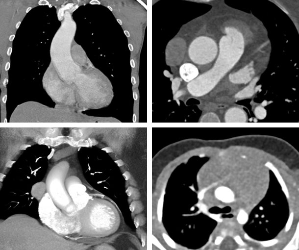
Chest CT Appearances
Thymoma CT Findings
- Typically 5-10cm in size
- Usually related to the superior pericardium that is anterior to the aorta, pulmonary artery, or superior vena cava (SVC)
- Usually well defined
- Round or lobulated
- Usually homogenous
- Enhances after contrast injection
- Often eccentric in location
- May look similar to lymphoma and teratoma
Related Pearls: Thymus