Chest CT Appearances
Septic Pulmonary Embolism (PE) CT Findings
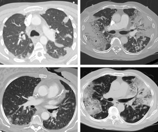
- Numerous bilateral nodules with varying degrees of cavitation (mostly in peripheral and lower lung fields)
- Infarcts may be seen concurrently in the kidneys, spleen or liver
- Wedge-shaped lesions with or without necrosis
- Air bronchograms within nodules
- Airspace opacities
Related Pearls: Pulmonary Embolism