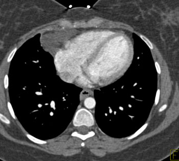
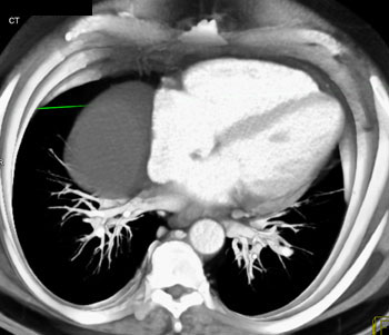
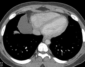
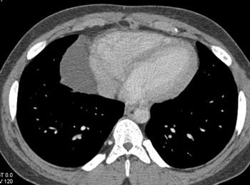
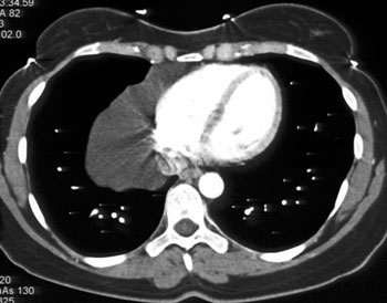
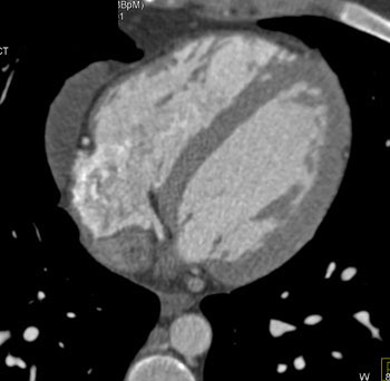
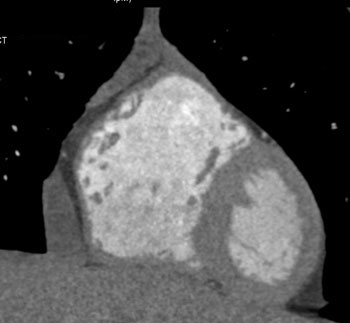
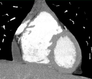
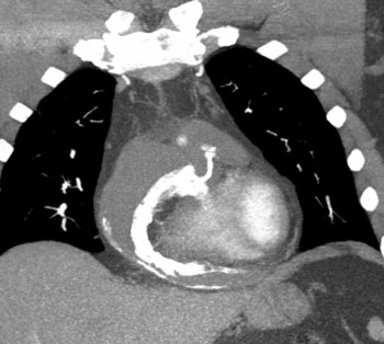
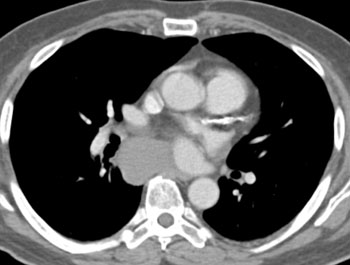
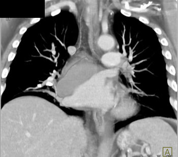
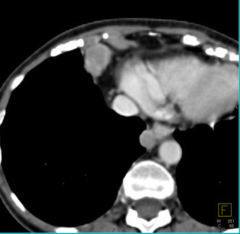
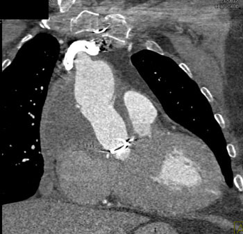
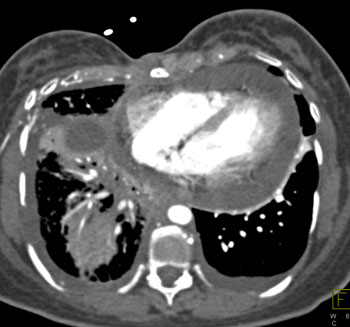
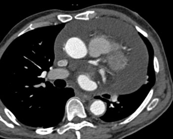
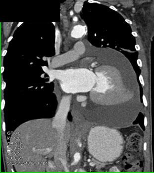
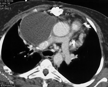
Diagnosis: Pericardial cyst Pericardial Cyst: Facts - Usually at right anterior costophrenic angle
- Water density on CT
- 2-30 cm in size
- Homogeneous without any enhancement
- Anomalous outpouching of the parietal pericardium
- Right costophrenic location in 70-90% of cases
- Size from 1 cm to >25 cm
- Usually incidental finding in an asymptomatic person
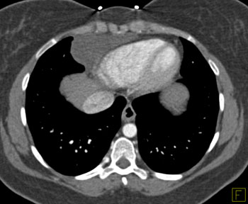
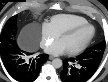
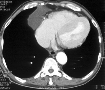
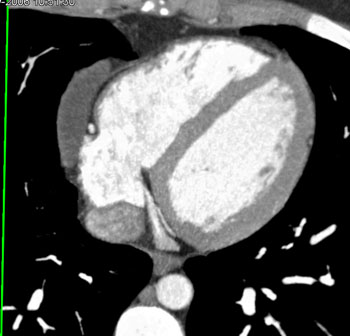
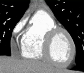
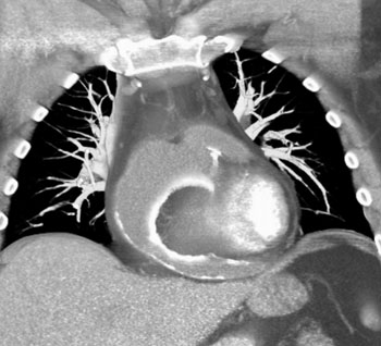
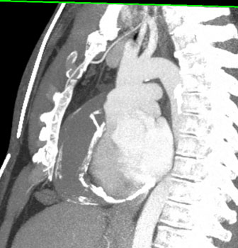
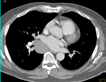
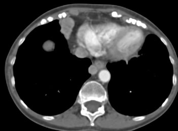
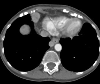
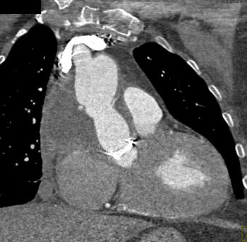
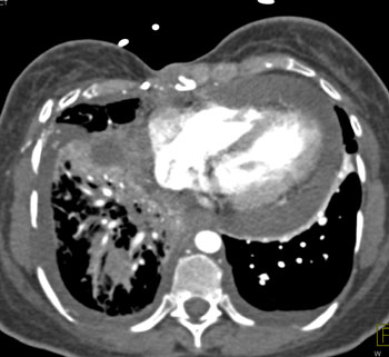
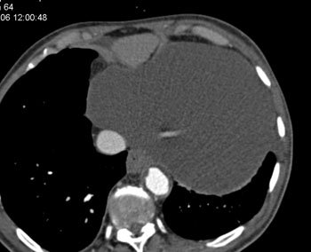
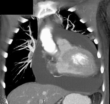
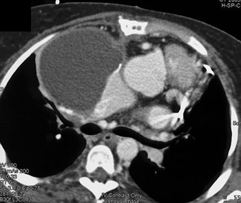
Pericardial Cysts: CT Findings- Smoothly marginated lesion surface
- Less than 10 HU
- No enhancement on contrast studies
- Size range from 2-30 cm
- Usually incidental finding in an asymptomatic person
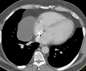
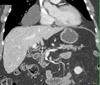
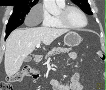
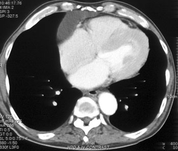
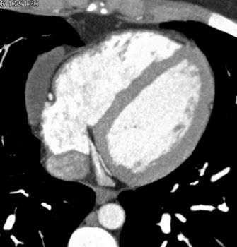
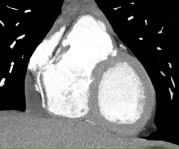
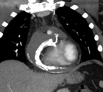
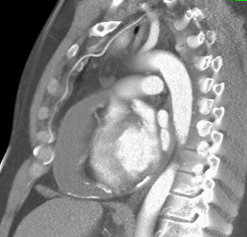
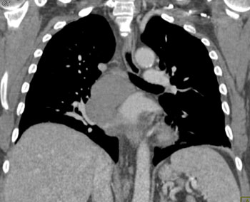
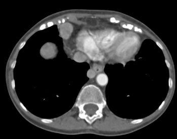
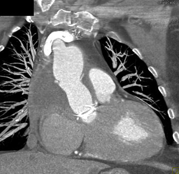
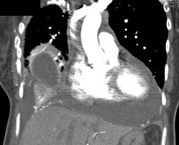
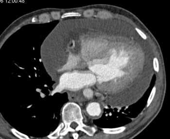
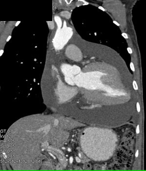
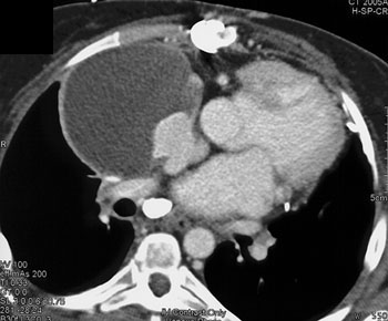
Pericardial Cyst: Facts- Usually an incidental finding
- May be confused with a Morgagni hernia or duplication cyst or occassionally a thymic cyst
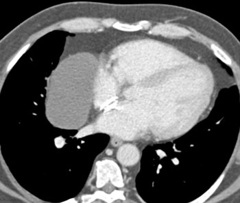
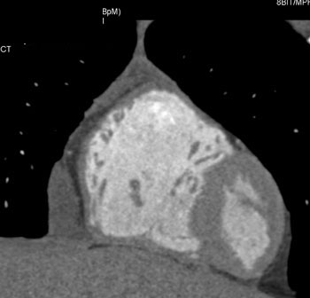
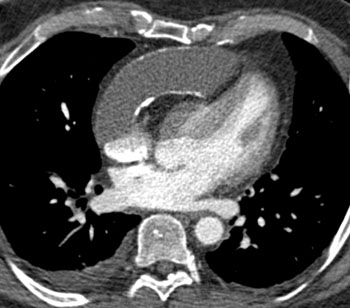
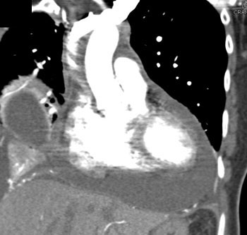
Pericardial cyst Pericardial Cyst: Facts - Usually at right anterior costophrenic angle
- Water density on CT
- 2-30 cm in size
- Homogeneous without any enhancement
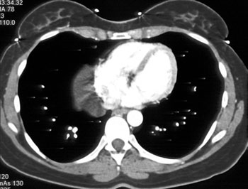
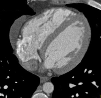
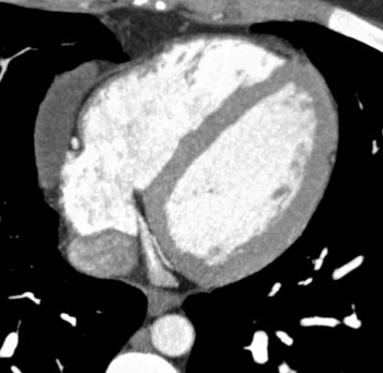
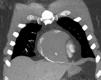
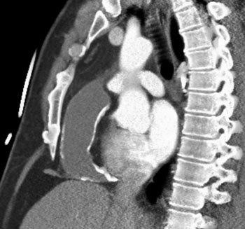
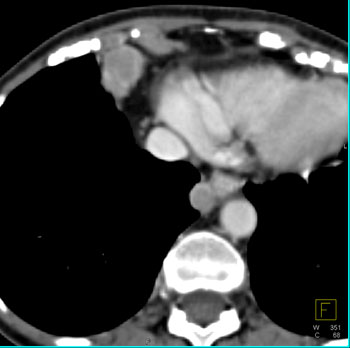
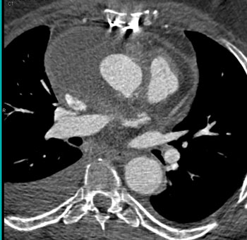
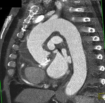
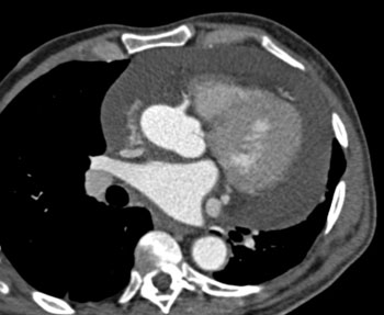
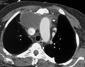
Pericardial Cyst Partially Calcified Pericardial Cyst Pericardial Cyst: Differential Diagnosis - Loculated pleural effusion
- Bronchogenic cyst
- Hematoma
- Esophageal duplication cyst
- Pericardial tumor
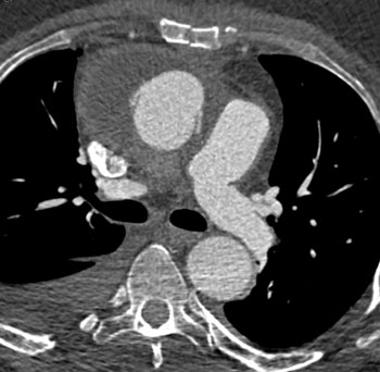
Pericardial Cysts Occassionally Can Be Confused With - Paracardiac nodes
- Loculated effusion
- Bronchogenic cyst
- Pericardial mass
- Loculated pericardial effusion
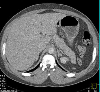
Bronchogenic Cyst Paracardiac Adenopathy Pericardial Effusion S/P AVR Pericardial Effusion Thymic cyst |