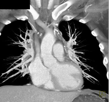
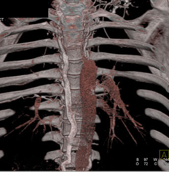
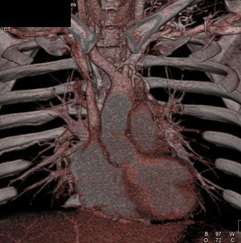
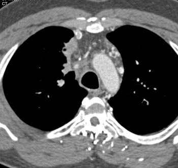
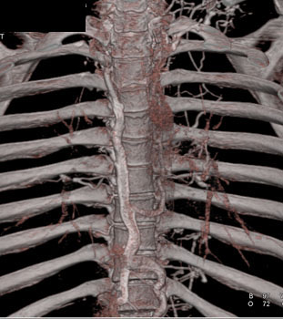
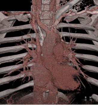
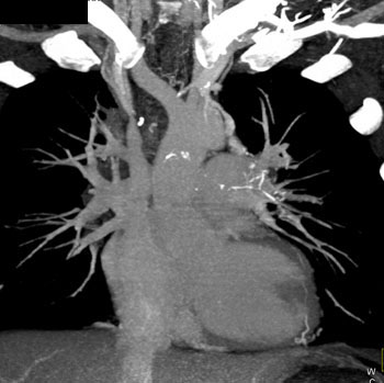
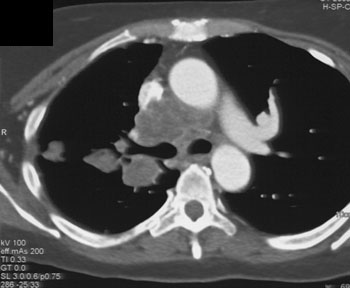
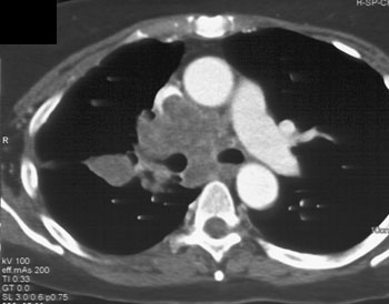
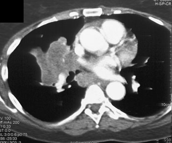
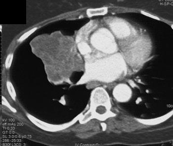
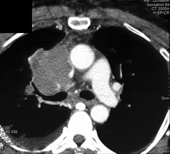
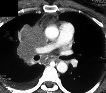
Diagnosis: Chronic SVC Occlusion with Extensive Mediastinal Collaterals SVC Obstruction: etiology - Lung cancer
- Lymphoma
- Thymoma
- Fibrosing mediastinitis (histoplasmosis is most common)
- Radiation fibrosis
- Iatrogenic (due to catheter placement)
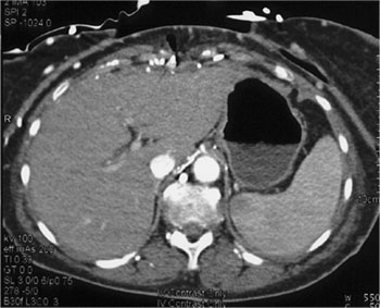
Collateral pathways are commonly opacified in SVC Syndrome - Azygous and hemiazygous system
- Paravertebral vessels
- Mediastinal veins as collaterals (as in this case)
- Anterior intercostal veins
- Internal mammary veins
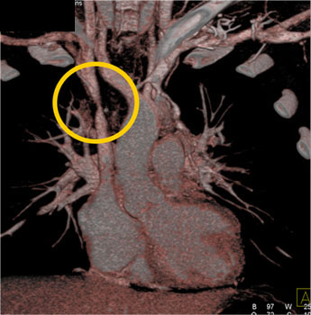
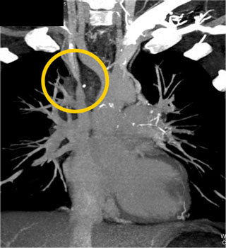
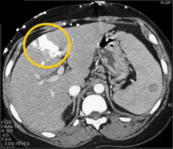
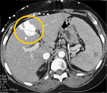
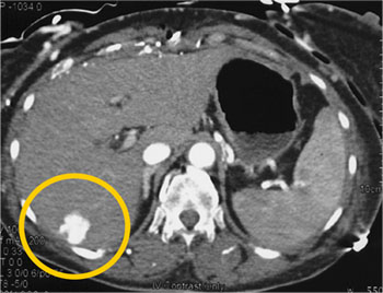
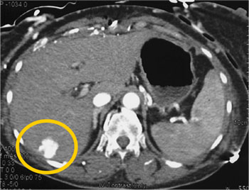
SVC Syndrome with Stenosis SVC Metastatic Renal Cell Carcinoma Invades the SVC SVC Encasement by Lung Cancer SVC Occlusion and "Hot Spot in the Liver" - Originally described on the nuclear medicine literature in the pre-CT days
- Can simulate a vascular metastases on early phase images of the liver (i.e. looks like a metastatic islet cell or neuroendocrine tumor)
- Will become isodense on delayed phase images
- Typical location near falciform ligament
- A key CT finding suggesting the diagnosis is collaterals in the subcutaneous tissues in the chest or upper abdomen
Hot Spot Liver Due to SVC Syndrome Collaterals Abdominal Wall and Hot Spot Liver |