- 2
- ,
- 3
- 8
- 1
To Quiz Yourself: Select OFF by clicking the button to hide the diagnosis & additional resources under the case.
Quick Browser: Select ON by clicking the button to hide the additional resources for faster case review.
CASE NUMBER
190


Diagnosis
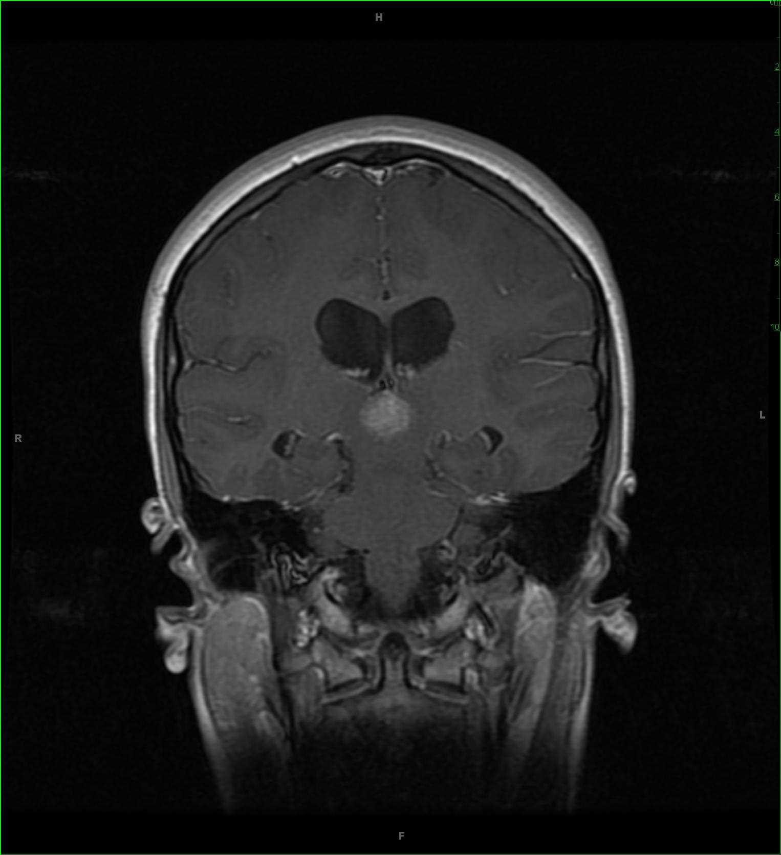
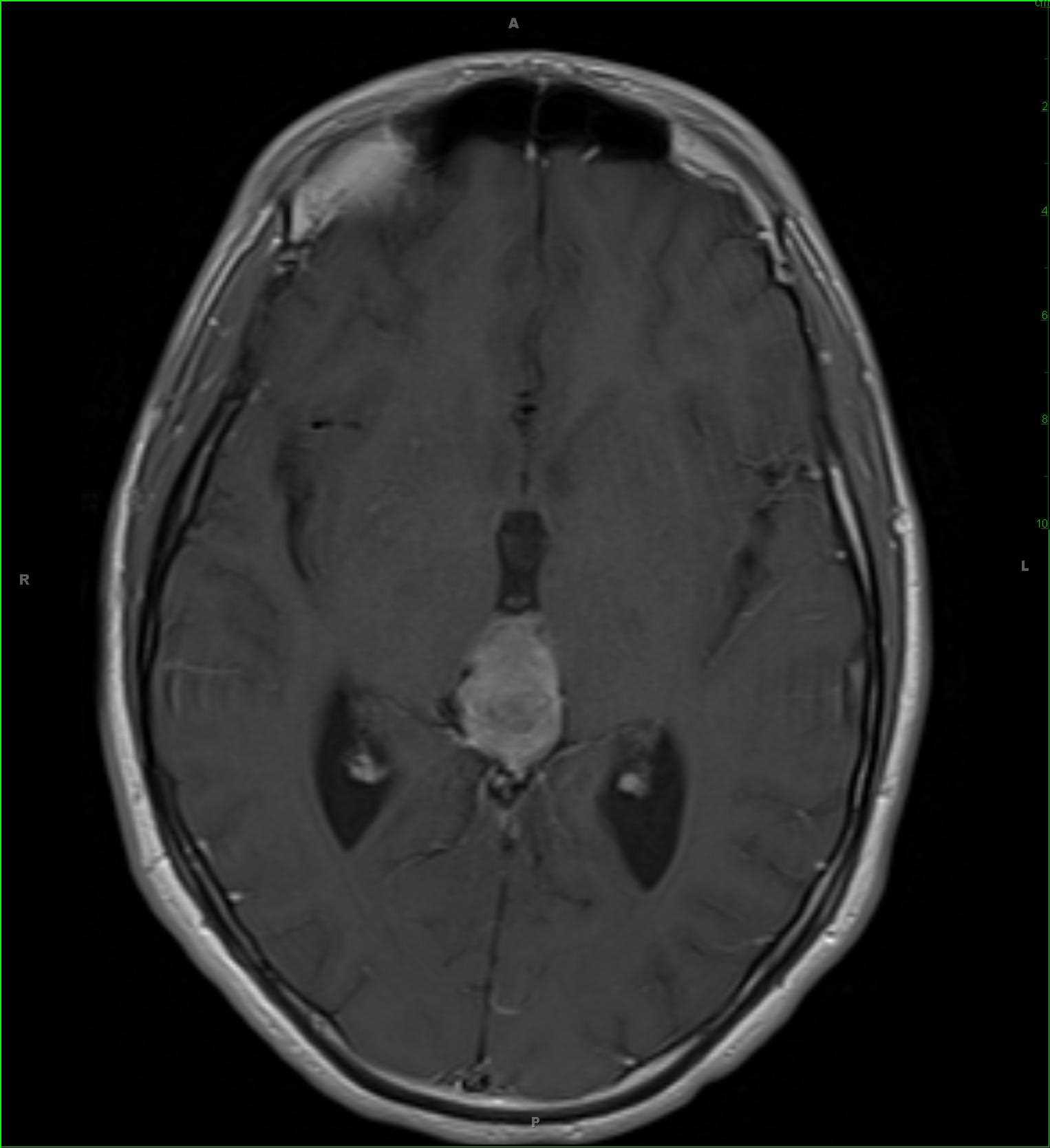
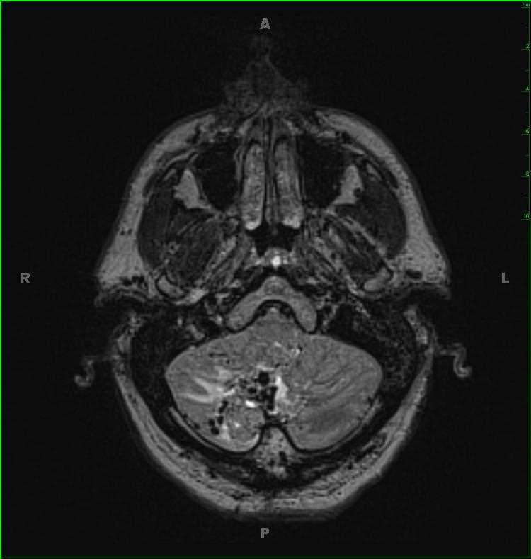
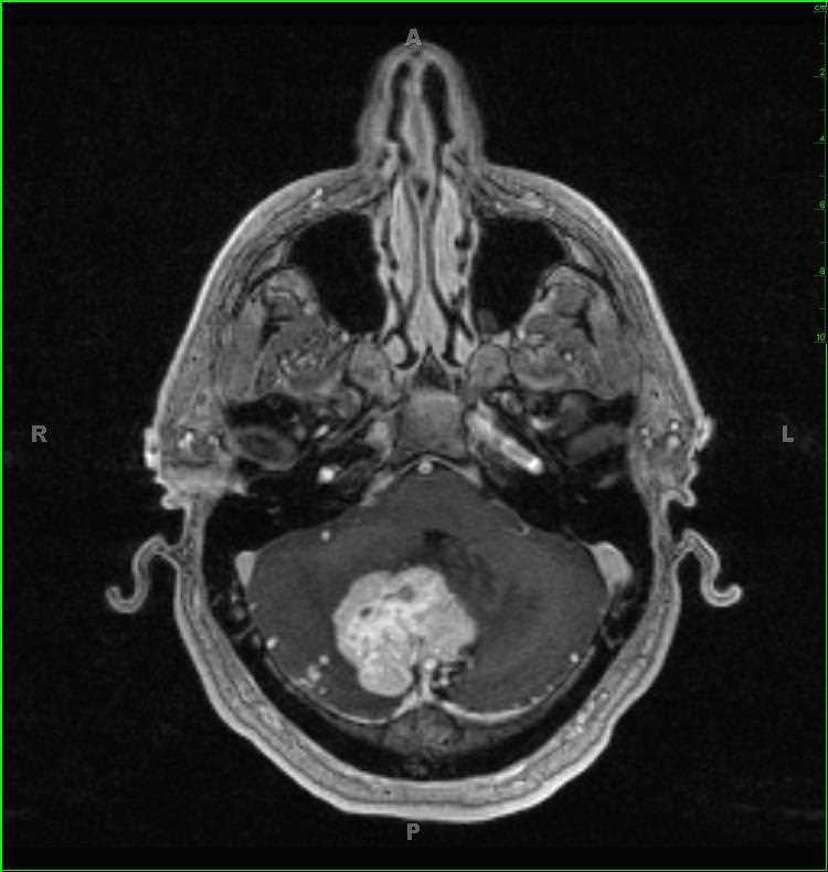
Hemangioblastoma
Note
52-year-old male who underwent a screening brain MRI for a family history of multiple pheochromocytomas and renal cysts. Poorly circumscribed, T1-hypointense mass which invades the cerebellar hemispheres and vermis. Several prominent flow voids are identified. There is significant peripheral vasogenic edema. The mass deforms the cerebellar hemispheres and vermis, resulting in effacement of the fourth ventricle. The mass enhances avidly and has increased blood volume. The findings are compatible with a hemangioblastoma, given the stated family history. Hemangioblastomas are most commonly found in cerebellum, brainstem, spinal cord. 25-40% of all hemangioblastomas occur in the setting of Von Hippel-Lindau syndrome. Primary treatment is with total resection.
Related videos to the case