Imaging Pearls ❯ Vascular ❯ Pulmonary Artery and PE
|
-- OR -- |
|
- “Pulmonary artery aneurysms and pseudoaneurysms are uncommon. Most are caused by trauma, often iatrogenic, infection, and Behçet’s syndrome. Less common causes include pulmonary hypertension, congenital heart disease, neoplasms, and connective tissue disease. Recognition of pulmonary artery aneurysms and pseudoaneurysms is important because of the high morbidity and mortality rates of rupture.”
Pulmonary artery aneurysms and pseudoaneurysms in adults: findings at CT and radiography.
Nguyen ET, Silva CI, Seely JM, Chong S, Lee KS, Müller NL.
AJR Am J Roentgenol. 2007 Feb;188(2):W126-34. - “By definition, an aneurysm is focal dilatationof a blood vessel that involves all three layers of vessel wall. A pseudoaneurysm does not involve all layers of the arterial wall and is therefore at higher risk of rupture. The upper limit of normal diameter of the main pulmonary artery on CT is 29 mm and of the right interlobar artery is 17 mm. We define an-eurysm as focal dilatation of a pulmonary artery beyond its maximal normal caliber.”
Pulmonary artery aneurysms and pseudoaneurysms in adults: findings at CT and radiography.
Nguyen ET, Silva CI, Seely JM, Chong S, Lee KS, Müller NL.
AJR Am J Roentgenol. 2007 Feb;188(2):W126-34. - “The most common forms of vasculitis as-sociated with pulmonary artery aneurysms are Behçet’s syndrome and Hughes-Stovin syndrome. Behçet’s syndrome is a chronic multisystem form of vasculitis characterized by recurrent oral and genital ulcers and uveitis. It is seen most commonly in Turkey and Southeast Asia. Behçet’s syndrome com-monly results in pulmonary artery aneurysms, which typically involve the right lower lobe arteries with frequent thrombosis and surrounding inflammation. Although these pulmonary artery aneurysms may regress with immunosuppressive medication, embolization is often needed to prevent life-threatening hemoptysis.”
Pulmonary artery aneurysms and pseudoaneurysms in adults: findings at CT and radiography.
Nguyen ET, Silva CI, Seely JM, Chong S, Lee KS, Müller NL.
AJR Am J Roentgenol. 2007 Feb;188(2):W126-34. - “Malpositioned Swan-Ganz catheters are an increasingly common cause of iatrogenic pulmonary artery pseudoaneurysm. In one prospective study with 500 consecutively enrolled patients, the incidence of rupture and hemorrhage after Swan-Ganz catheter insertiowas 0.2%. The complication occurs mainly inpatients in whom the Swan-Ganz catheter has been inserted too far into a pulmonary arterial branch. The tip of the catheter begins to erode the wall of the artery and causes weakening and dilatation. The vessel ruptures where extravasated blood is contained by adventitia, or thrombus forms a pseudoaneurysm .”
Pulmonary artery aneurysms and pseudoaneurysms in adults: findings at CT and radiography.
Nguyen ET, Silva CI, Seely JM, Chong S, Lee KS, Müller NL.
AJR Am J Roentgenol. 2007 Feb;188(2):W126-34. - “Intrinsic weakness in the arterial wall dueto connective tissue abnormalities such as Marfan syndrome, Ehlers-Danlos syndrome, and cystic medial necrosis also predispose to aneurysm formation. Aneurysms in these pa-tients typically involve the aorta but also can affect the pulmonary arteries.”
Pulmonary artery aneurysms and pseudoaneurysms in adults: findings at CT and radiography.
Nguyen ET, Silva CI, Seely JM, Chong S, Lee KS, Müller NL.
AJR Am J Roentgenol. 2007 Feb;188(2):W126-34. - “Although pulmonary artery aneurysms and pseudoaneurysms are uncommon, knowledge of their congenital and acquired causes and radiologic manifestations is important. Assessment with contrast-en-hanced CT allows accurate evaluation of pulmonary artery aneurysms and pseudo-aneurysms, facilitating prompt diagnosis and treatment.”
Pulmonary artery aneurysms and pseudoaneurysms in adults: findings at CT and radiography.
Nguyen ET, Silva CI, Seely JM, Chong S, Lee KS, Müller NL.
AJR Am J Roentgenol. 2007 Feb;188(2):W126-34.
- “Pulmonary artery (PA) aneurysms (PAAs) are rare and infrequently diagnosed. Deterling and Clagett1 discovered 8 cases of PAAs in 109 571 consecutive postmortem examinations. PAAs generally occurred in a younger age group than aortic aneurysms with an equal sex incidence.2 Eighty-nine percent of all PAAs were located in the main PA, whereas only 11% were located in the pulmonary branches.3 When affecting the PA branches, PAAs in the left PA were more common than in the right PA.”
Aneurysms of the Pulmonary Artery
Maximilian Kreibich, et al.
Circulation Volume 131, Issue 3, 20 January 2015; Pages 310-316 - Causes of Pulmonary Artery Aneurysms
Connective tissue abnormalities
- Ehlers-Danlos syndrome
- Marfan syndrome
- Cystic medial necrosis
Infectious
- Syphilis
- Tuberculosis
- Pyogenic bacteria
- Septic embolisms
- Bacterial and fungal pneumonia - Causes of Pulmonary Artery Aneurysms
Vasculitis
- Behçet syndrome
- Hughes-Stovin syndrome
Heart defects
- Persistent ductus arteriosus
- Ventricular septal defects
- Atrial septal defects
- Hypoplastic aortic valve
- Bicuspid aortic valve
- Pulmonary valve stenosis
- Pulmonary regurgitation
- Absent pulmonary valve
- “The true mortality associated with undiagnosed pulmonary embolism is estimated to be less than 5%, but recovery from pulmonary embolism is associated with complications such as bleeding due to anticoagulant treatment, recurrent venous thromboembolism, chronic thromboembolic pulmonary hypertension, and long-term psychological distress. Approximately half the patients who receive a diagnosis of pulmonary embolism have functional and exercise limitations 1 year later (known as post–pulmonary-embolism syndrome), and the health-related quality of life for patients with a history of pulmonary embolism is diminished as compared with that of matched controls. Therefore, the timely diagnosis and expert management of pulmonary embolism are important.”
Pulmonary Embolism
Susan R. Kahn, Kerstin de Wit
N Engl J Med 2022;387:45-57. 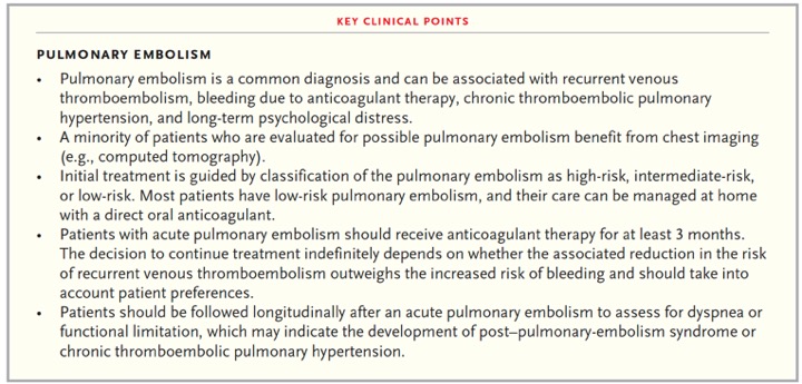
Pulmonary Embolism
Susan R. Kahn, Kerstin de Wit
N Engl J Med 2022;387:45-57.- • Pulmonary embolism is a common diagnosis and can be associated with recurrent venous thromboembolism, bleeding due to anticoagulant therapy, chronic thromboembolic pulmonary hypertension, and long-term psychological distress.
• A minority of patients who are evaluated for possible pulmonary embolism benefit from chest imaging (e.g., computed tomography).
• Initial treatment is guided by classification of the pulmonary embolism as high-risk, intermediate-risk, or low-risk. Most patients have low-risk pulmonary embolism, and their care can be managed at home with a direct oral anticoagulant.
Pulmonary Embolism
Susan R. Kahn, Kerstin de Wit
N Engl J Med 2022;387:45-57. - • Patients with acute pulmonary embolism should receive anticoagulant therapy for at least 3 months. The decision to continue treatment indefinitely depends on whether the associated reduction in the risk of recurrent venous thromboembolism outweighs the increased risk of bleeding and should take into account patient preferences.
• Patients should be followed longitudinally after an acute pulmonary embolism to assess for dyspnea or functional limitation, which may indicate the development of post–pulmonary-embolism syndrome or chronic thromboembolic pulmonary hypertension.
Pulmonary Embolism
Susan R. Kahn, Kerstin de Wit
N Engl J Med 2022;387:45-57. - “Occult cancer is detected in 5.2% of patients within 1 year after a diagnosis of unprovoked pulmonary embolism. An extensive screening strategy may detect more cancers than limited screening, but data are limited as to whether such screening is associated with better patient outcomes. Experts recommend limited cancer screening guided by medical history, physical examination, basic laboratory tests and chest radiographs, and age-specific and sex-specific cancer screening.”
Pulmonary Embolism
Susan R. Kahn, Kerstin de Wit
N Engl J Med 2022;387:45-57. - "Appropriate management of subsegmental pulmonary embolism (a single isolated subsegmental pulmonary embolus or multiple emboli, without the presence of pulmonary embolism in segmental or more proximal pulmonary vessels and without deep-vein thrombosis in the legs) is uncertain. Although some guidelines suggest clinical surveillance instead of anticoagulation in patients with low-risk subsegmental pulmonary embolism, a recent prospective cohort study involving such patients who were treated without anticoagulation therapy showed a higher-than expected incidence of recurrent venous thromboembolism during 90-day follow-up A randomized, placebo-controlled trial of clinical surveillance as compared with anticoagulation in this patient population is ongoing (ClinicalTrials.gov number, NCT04263038).”
Pulmonary Embolism
Susan R. Kahn, Kerstin de Wit
N Engl J Med 2022;387:45-57.

- Pulmonary Artery Aneurysm: Causes
• Congenital (50%)
• In general, it is presumed that increased flow caused by left-to-right shunt results in increased hemodynamic shear stress on the vessel walls and therefore promotes aneurysm formation in the PAs.The 3 most frequent congenital heart defects associated with a PAA are, in decreasing order, persistent ductus arteriosus, ventricular septal defects, and atrial septal defects. - Pulmonary Artery Aneurysm: Causes
• Congenital (50%)
• Pulmonary valve stenosis
• Ehlers-Danlos syndrome
• Marfan syndrome
• Cystic medial necrosis
• Untreated syphilis and tuberculosis
• IVDA
• Vasculitis (Behcet Syndrome) - “In general, clinical manifestations of PAA remain nonspecific, whereas most patients with a PAA, even those with large PAA diameters up to 70 mm, have no complaints.Clinical symptoms include dyspnea, chest pain, hoarseness, palpitation, and syncopal episodes. Bronchus compression by a large PAA may be responsible for cyanosis, cough, and increasing dyspnea, pneumonia, fever, and bronchiectasis. In addition, patients with PAA have a high incidence of pulmonary emboli.”
Aneurysms of the Pulmonary Artery Maximilian Kreibich et al. Circulation. 2015;131:310-316 - “Overall, surgery remains the cornerstone of therapy for lesions involving the main pulmonary trunk, and evidence suggesting an absolute diameter threshold for surgery of the main PA is lacking. However, from our clinical experience and scientific knowledge of all the available data about aortic aneurysms, we suggest operating on adults with pulmonary trunk aneurysms >5.5 cm according to the guidelines for aortic disease.”
Aneurysms of the Pulmonary Artery Maximilian Kreibich et al. Circulation. 2015;131:310-316 - “In case of conservative treatment, it is our opinion that patients should be re-evaluated regularly, and a change in treatment should strongly be considered in case of compression of adjacent structures, thrombus formation in the aneurysm sack, ≥5-mm increase in the diameter of the aneurysm in 6 months, the appearance of clinical symptoms, evidence of valvular pathologies or shunt flow, and verification of PAH.”
Aneurysms of the Pulmonary Artery Maximilian Kreibich et al. Circulation. 2015;131:310-316
- “The qualities of pulmonary CTA make it desirable to clinicians as a test for suspected PE. The apparent binary yes or no answer to a challenging diagnosis with ambiguous clinical presentation is enticing. As with other diagnostic tests, however, the posttest probability of pulmonary CTA hinges on pretest clinical assessment. Validated clinical decision rules and integrated CDS systems can influence the appropriate use of pulmonary CTA, but further investigation is required to de ne the most successful means of integration into clinical practice.”
Role of Clinical Decision Tools in the Diagnosis of Pulmonary Embolism William M. Sherk, Jadranka Stojanovska AJR 2017; 208:60–70 - “Single or limited educational interventions to inform clinicians of evidence-based decision rules do not overcome the obstacles. Repetitive educational efforts over time—possibly as long as 5 years—in- uence change. Computerized forms of CDS facilitate tracking of individual clinician use and yield of pulmonary CTA and in turn al- low opportunities for feedback. Feedback systems integrated into the electronic medical record show potential for promoting adherence to evidence-based guidelines and use of clinical decision rules before ordering of pulmonary CTA .”
Role of Clinical Decision Tools in the Diagnosis of Pulmonary Embolism William M. Sherk, Jadranka Stojanovska AJR 2017; 208:60–70
- “The largest meta-analysis to date exam- ined over 10,000 patients up to the year 2009 and reported incidental PE had a prevalence of 2.6% (95% CI, 1.9–3.4%), with a higher prevalence in patients with VTE risk factors such as malignancy (3.1%) and inpatient status (4.0%).”
Management of the Incidental Pulmonary Embolism Victor Chiu, Casey O’Connell AJR 2017; 208:485–488 - “Although treatment of symptomatic PE with anticoagulation results in a clear reduction in mortality, the bene ts of treating incidental PE have not yet been evaluated in a large prospective study. The risk of major bleeding while on anticoagulation for any VTE is 7.2 per 100 patient-years, making the decision to treat one of great consequence, particularly in higher risk patients such as those in the ICU and those with cancer.”
Management of the Incidental Pulmonary Embolism Victor Chiu, Casey O’Connell AJR 2017; 208:485–488 - “The National Comprehensive Cancer Network also recommends treatment of incidental PE similar to that for symptomatic PE in patients with cancer and recommends against routinely obtaining repeat imaging.”
Management of the Incidental Pulmonary Embolism Victor Chiu, Casey O’Connell AJR 2017; 208:485–488 - “Pulmonary CTA is well established as a fast and reliable means of excluding or diagnosing PE. Continued developments in CT system hardware and postprocessing techniques will allow incremental reductions in radiation and contrast material requirements while improving image quality. Advances in risk strati cation and prognostication from pulmonary CTA examinations should further re ne its clinical value while minimizing the potential harm from overutilization and overdiagnosis."
State-of-the-Art Pulmonary CT Angiography for Acute Pulmonary Embolism Moritz H. Albrecht et al. AJR 2017; 208:495–504 - “This technique is effective despite the variable embrace by clinicians of the d-dimer test, a test that suffers from low specificity. However, the advantage of this laboratory marker lies in its high negative predictive value, so that acute PE can be safely excluded by a negative d-dimer result. In case of elevated d-dimer values, pulmonary CTA should be performed."
State-of-the-Art Pulmonary CT Angiography for Acute Pulmonary Embolism Moritz H. Albrecht et al. AJR 2017; 208:495–504 - “For example, pulmonary CTA has emerged as a formidable prognostic marker to gauge the
severity of hemodynamic compromise from acute PE and identify patients at heightened risk for fatal or nonfatal adverse events, thus guiding clinical management toward more aggressive therapy. The main methods that have been described to categorize the hemodynamic relevance and severity of PE are imaging markers of right heart strain, methods for clot burden quantification, and lung perfusion measurements."
State-of-the-Art Pulmonary CT Angiography for Acute Pulmonary Embolism Moritz H. Albrecht et al. AJR 2017; 208:495–504 - “For clinical purposes, across all endpoints, the right ventricle (RV) diameter–left ventricle (LV) diameter ratio on pulmonary CTA has the strongest predictive value and most robust evidence base for adverse clin- ical outcomes in patients with acute PE. A ratio of more than 1 on traverse images and of more than 0.9 using true four-chamber view reconstructions is considered indicative of right heart strain and has been shown to predict adverse outcomes and early death."
State-of-the-Art Pulmonary CT Angiography for Acute Pulmonary Embolism Moritz H. Albrecht et al. AJR 2017; 208:495–504 - “One study investigating spectral optimization in monochromatic dual-energy pulmonary CTA with reduced iodine load suggested that 60 keV may be the optimal energy level to analyze the thoracic circulation . Other investigators have also concluded that iodine load can be reduced when virtual monoenergetic images extrapolated to photon energies of 50 or 70 keV are used ."
State-of-the-Art Pulmonary CT Angiography for Acute Pulmonary Embolism Moritz H. Albrecht et al. AJR 2017; 208:495–504 - “Pulmonary CTA is well established as a fast and reliable means of excluding or diagnosing PE. Continued developments in CT system hardware and postprocessing techniques will allow incremental reductions in radiation and contrast material requirements while improving image quality. Advances in risk stratification and prognostication from pulmonary CTA examinations should further refine its clinical value while minimizing the potential harm from overutilization and overdiagnosis."
State-of-the-Art Pulmonary CT Angiography for Acute Pulmonary Embolism Moritz H. Albrecht et al. AJR 2017; 208:495–504
- “The incidence of PE is highest in the inpatient setting. While the incidence of PE in outpatient and emergency department patients is similar, more CTA thorax examinations are ordered in the emergency department than in the outpatient setting. There is no difference in the incidence of PE based on who orders CTA thorax examinations.”
Incidence of pulmonary emboli on chest computed tomography angiography based upon referral patterns Meesa IR et al. Emerg Radiol (2016) 23::251–254 - “Our study shows that there is no statistical difference in the incidence of PE when the studies are ordered by attending physicians, residents, or physician extenders. The reasoning might be that in many training institutions, including ours, the attending physician often supervises the residents and physician extenders and, therefore, is often involved in the decision-making process of whether or not to order a CTA to exclude PE. We are somewhat limited in making this conclusion because there are no similar studies in the literature to compare our results against or a way to confirm using the EMR and a retrospective methodology.”
Incidence of pulmonary emboli on chest computed tomography angiography based upon referral patterns Meesa IR et al. Emerg Radiol (2016) 23::251–254 - “Our study indicates an incidence of PE in inpatients of 19.2 %, in ED patients of 6.7 %, and outpatients of 6.4 %. This compares to Mamlouk where the incidences were 13.46 % in inpatients and 6.36 % in ED patients. The large adult sample size of our study resulted in high confidence levels. ”
Incidence of pulmonary emboli on chest computed tomography angiography based upon referral patterns Meesa IR et al. Emerg Radiol (2016) 23::251–254 - “The SCAs may manifest a wide spectrum of pathologic conditions ranging from common atherosclerosis to more rare entities such as vasculitis. MDCT angiography performed using proper technique is an excellent modality for the diagnosis, follow-up, and treatment planning of a wide range of disease processes involving the SCAs. Knowledge of the typical MDCT findings and of potential complications allows the radiologist to make a timely and accurate diagnosis.”
Nontraumatic Subclavian Artery Abnormalities: Spectrum of MDCT Findings
Jones CS, Verde F, Johnson PT, Fishman EK. AJR Am J Roentgenol. 2016 May 17:1-8
- “In this rare vascular developmental anomaly, the left pulmonary artery arises from the posterior aspect of the right pulmonary artery and passes between the trachea and esophagus to reach the left hilum. The left pulmonary artery thus forms a sling around the distal trachea and the proximal right main bronchus.”
Congenital and Acquired Pulmonary Artery Anomalies in the Adult: Radiologic Overview Castañer E et al. RadioGraphics 2006 26:2, 349-371 - “Those affected by pulmonary artery sling may be classified generally into two groups: one with a normal bronchial pattern and the other with one or more malformations of the bronchotracheal tree (eg, stenosis of a long segment of the trachea or absence of the pars membranacea) as well as cardiovascular abnormalities. In the latter group, mortality and morbidity are high during infancy.The former group includes very few asymptomatic adults. In asymptomatic cases, a pulmonary artery sling may mimic a mediastinal mass on chest radiographs. CT and magnetic resonance (MR) imaging may be used to establish the diagnosis with certainty.”
Congenital and Acquired Pulmonary Artery Anomalies in the Adult: Radiologic Overview Castañer E et al. RadioGraphics 2006 26:2, 349-371 - “Idiopathic dilatation of the pulmonary trunk is a rare congenital anomaly that involves abnormal enlargement of the pulmonary trunk, with or without dilatation of the right and left pulmonary arteries . To reach this diagnosis, it is necessary to exclude pulmonary and cardiac diseases (mainly pulmonary valve stenosis) and to confirm the presence of normal pressure in the right ventricle and pulmonary artery.”
Congenital and Acquired Pulmonary Artery Anomalies in the Adult: Radiologic Overview Castañer E et al. RadioGraphics 2006 26:2, 349-371 - “Aneurysms or pseudoaneurysms of the pulmonary arteries, whether congenital or acquired, are rare. They may occur in association with a congenital cardiovascular anomaly, especially patent ductus arteriosus; infection (mycotic aneurysm); trauma, most commonly as a result of pulmonary artery perforation (due to improper placement of a catheter) or penetrating injury and very rarely as a result of blunt injury; vascular abnormality (eg, cystic medial necrosis, Behçet disease, Marfan syndrome, and Takayasu disease); and pulmonary hypertension.”
Congenital and Acquired Pulmonary Artery Anomalies in the Adult: Radiologic Overview Castañer E et al. RadioGraphics 2006 26:2, 349-371 - “Most mycotic aneurysms are secondary to endovascular seeding due to septic pulmonary emboli and are found in patients with endocarditis. Aneurysms secondary to a direct extension of infection from the adjacent parenchyma are seen in patients with necrotizing pneumonia or chronic tuberculosis. Mycotic aneurysms can be single or multiple and can be located centrally or peripherally.”
Congenital and Acquired Pulmonary Artery Anomalies in the Adult: Radiologic Overview Castañer E et al. RadioGraphics 2006 26:2, 349-371 - “Rasmussen aneurysm is a rare condition caused by weakening of the pulmonary artery wall from adjacent cavitary tuberculosis. Hemoptysis is the usual symptom at initial manifestation. Although the source of hemoptysis in cavitary tuberculosis is usually the bronchial arteries, Rasmussen aneurysm usually occurs in a peripheral pulmonary artery (. Chest radiographic findings that may suggest the formation of a pseudoaneurysm include an intracavitary protrusion, the replacement of a cavity by a nodule, or a rapidly growing mass.”
Congenital and Acquired Pulmonary Artery Anomalies in the Adult: Radiologic Overview Castañer E et al. RadioGraphics 2006 26:2, 349-371
- "Practical measures to reduce the risk of PE misdiagnosis could and should include any of the following: systematic use of pretest probability assessment (which would require buy-in from clinicians and incorporation into imaging protocols); radiology technologists being educated to optimize image quality, focusing on proper patient breathing technique and repeating examinations where appropriate; increased familiarization by radiologists with the range of potential diag- nostic pitfalls; encouragement of the use of second opinions by interpreting radiologists, particularly for solitary subsegmental PEs; and regular review of positive pulmonary CTA cases (e.g., at monthly discrepancy or audit meetings). Some of these measures are easier to implement than others, but their importance is underscored by the implications of a false-positive diagnosis of PE.”
Overdiagnosis of Pulmonary Embolism by Pulmonary CT Angiography Hutchinson BD et al. AJR 2015; 205:271–277 - "Practical measures to reduce the risk of PE misdiagnosis could and should include any of the following: systematic use of pretest probability assessment (which would require buy-in from clinicians and incorporation into imaging protocols); radiology technologists being educated to optimize image quality, focusing on proper patient breathing technique and repeating examinations where appropriate; increased familiarization by radiologists with the range of potential diag- nostic pitfalls; encouragement of the use of second opinions by interpreting radiologists, particularly for solitary subsegmental PEs; and regular review of positive pulmonary CTA cases (e.g., at monthly discrepancy or audit meetings).”
Overdiagnosis of Pulmonary Embolism by Pulmonary CT Angiography Hutchinson BD et al. AJR 2015; 205:271–277 - “In routine clinical practice, PEs diagnosed by pulmonary CTA are frequently overdiagnosed, when compared with the consensus opinion of a panel of expert chest radiologists. Improvements in the quality of pulmonary CTA examination and increased fa- miliarity with potential diagnostic pitfalls in pulmonary CTA are recommended to minimize misdiagnosis of PE.”
Overdiagnosis of Pulmonary Embolism by Pulmonary CT Angiography Hutchinson BD et al. AJR 2015; 205:271–277 - “A total of 937 pulmonary CTA studies were performed over the study period. PE was diagnosed in the initial report in 174 of these cases (18.6%). There was discordance between the chest radiologists and the original radiologist in 45 of 174 (25.9%) cases. Discordance occurred more often where the original reported PE was solitary (46.2% of reported solitary PEs were considered negative on retrospective review) and located in a segmental or subsegmental pulmonary artery (26.8% of segmental and 59.4% of subsegmental PE diagnoses were considered negative on retrospective review). The most common cause of diagnostic
difficulty was breathing motion artifact, followed by beam-hardening artifact.”
Overdiagnosis of Pulmonary Embolism by Pulmonary CT Angiography Hutchinson BD et al. AJR 2015; 205:271–277 - “The risk of hemorrhage related to anticoagulation therapy is potentially significant. A large meta-analysis in 2003 [8] found a 7% annual risk of major bleeding and a 0.4% incidence of bleeding-related fatality in pa- tients treated with oral anticoagulation therapy for venous thromboembolism for longer than 3 months. The practical implications of long-term anticoagulation therapy for the patient are also potentially significant, requir- ing frequent attendance to their medical practitioners for blood tests, consequent time off from work, potential adverse drug interactions with other medications, adjustments to travel and lifestyle, implications for future dental and medical procedures, and possible negative effects on life insurance status.”
Overdiagnosis of Pulmonary Embolism by Pulmonary CT Angiography Hutchinson BD et al. AJR 2015; 205:271–277 - “This study shows an unexpectedly high rate of overdiagnosis of PE by pulmonary CTA in a tertiary-care university hospital, with an overall rate of 25.9% of all positive pulmonary CTA examinations, increasing to as high as 66.7% of cases where a solitary subsegmental PE was originally reported. Discordance was greatest for solitary PEs, PEs located in segmental and subsegmental pulmonary arteries, and in the lower zones of the lungs. The positive predictive value of pulmonary CTA for the diagnosis of PE was only 74.1% in this study.”
Overdiagnosis of Pulmonary Embolism by Pulmonary CT Angiography Hutchinson BD et al. AJR 2015; 205:271–277
- “Vascular anomalies are one important component that can be discovered incidentally on imaging. Early detection of the asymptomatic early process that could be managed with early intervention is paramount, particularly for aortic aneurysm management. We identified incidental abdominal aortic aneurysms requiring monitoring or surgical treatment in five cases.”
Incidental Findings at Initial Imaging Workup of Patients with Prostate Cancer: Clinical Signiificance
Elmi A et al.
AJR 2012; 199:1305-1311 - “ Septic pulmonary embolism (SPE) is an uncommon disorder that generally presents with an insidious onset of fever, respiratory symptoms, and lung infiltrates. Clinical and radiologic features at presentation are usually nonspecific, and the diagnosis of this disorder is frequently delayed. Historically, SPE has been associated with risk factors such as IV drug use, pelvic thrombophlebitis, and suppurative processes in the head and neck.However, increasing use of indwelling catheters and devices as well as increasing numbers of immunocompromised patients have changed the epidemiology and clinical manifestations of SPE.”
Septic Pulmonary Embolism: Presenting Features and Clinical Course of 14 Patients
Cook RJ et al.
Chest 2005;128(1):162-166 - “Historically, SPE has been associated with risk factors such as IV drug use, pelvic thrombophlebitis, and suppurative processes in the head and neck.However, increasing use of indwelling catheters and devices as well as increasing numbers of immunocompromised patients have changed the epidemiology and clinical manifestations of SPE.”
Septic Pulmonary Embolism: Presenting Features and Clinical Course of 14 Patients
Cook RJ et al.
Chest 2005;128(1):162-166 - “ Septic pulmonary embolism (SPE) is an uncommon disorder that generally presents with an insidious onset of fever, respiratory symptoms, and lung infiltrates. Clinical and radiologic features at presentation are usually nonspecific, and the diagnosis of this disorder is frequently delayed.”
Septic Pulmonary Embolism: Presenting Features and Clinical Course of 14 Patients
Cook RJ et al.
Chest 2005;128(1):162-166 - “Although findings on CXR tend to be nonspecific, CT may yield helpful clues that may suggest the diagnosis of SPE. Parenchymal lesions related to SPE are usually multiple and nodular with a peripheral distribution and a tendency for cavitation. These features associated with an extrapulmonary focus of infection should lead to consideration of SPE as the cause. Although other authors have described a “feeding vessel” sign (a vessel leading to a peripheral lung lesion) as a characteristic feature of SPE, we were able to identify this feature associated with only a minority of parenchymal lesions and did not find it particularly helpful in the recognition of SPE.”
Septic Pulmonary Embolism: Presenting Features and Clinical Course of 14 Patients
Cook RJ et al.
Chest 2005;128(1):162-166
- "As in MDCT with a smaller number of slices, the combination of CTV with CTPA in 64-MDCT results in a small but definite increase in the percentage of patients with the diagnosis of thromboembolic disease."
64-MDCT Pulmonary Angiography and CT Venography in the Diagnosis of Thromboembolic Disease
Nazaroglu H et al.
AJR 2009; 192:654-661 - "CTV started 210 seconds after the start of the contrast injection. The region from the popliteal fossa to 3-4 cm proximal to the iliac crest was scanned helically from a caudal to cranial direction."
64-MDCT Pulmonary Angiography and CT Venography in the Diagnosis of Thromboembolic Disease
Nazaroglu H et al.
AJR 2009; 192:654-661 - Pulmonary Artery Pseudoaneurysm: Clinical Presentation
- Hemoptysis (which may be brisk)
- Mass or rounded infiltrate on CXR
- Opacification of hemi-thorax on CXR or CT scan
- Management must be aggressive as 100% mortality with rupture - Pulmonary Artery Pseudoaneurysm: Causes Vascular abnormality
- Behcet disease
- Marfan syndrome
- Takayasu disease
Other causes
- Septic emboli
- Neoplasm
- Pulmonary Artery Pseudoaneurysm: Causes Infection
- Mycotic aneurysm (direct extension from necrotizing pneumonia or endovascular seeding from endocarditis)
- Mycobacterial aneurysm (Rasmussen Aneurysm)
Trauma
- Improper insertion of Swan Ganz
- Penetrating trauma
- "The daily use of MDCT studies for the evaluation of pulmonary embolic disease or aortic abnormalities can reveal incidental PDAs. Small incidental PDAs can be identified on chest MDCT angiography timed for either the pulmonary arteries or the aorta."
Incidental Finding on MDCT of Patent Ductus Arteriosus: Use of CT and MRI to Assess Clinical Importance
Goitein O et al.
AJR 2005;184:1924-1931
- "Our data showed a suboptimal use of the Wells criteria and subjective overestimation of the probability of PE before ordering of CTA. Although a definitive acceptable PE positivity rate for CTA has not been established, the 10% yield represents overuse of CTA as a screening rather than a diagnostic examination."
CT Angiography in the Evaluation of Acute Pulmonary Embolus
Costantion MM et al.
AJR 2008; 191:471-474
- "More than 80% of deaths from PE occurring in the first 30 minutes, and 90% within the first 2.5 hours of the event."
CT Angiography in the Evaluation of Acute Pulmonary Embolus
Costantion MM et al.
AJR 2008; 191:471-474
- "The patients who survive to be referred for diagnostic evaluation are a very different subset of this population. It has been suggested that the mortality and recurrence rates in this population are likely as low as 5%, even if the patient is not treated ."
CT Angiography in the Evaluation of Acute Pulmonary Embolus
Costantion MM et al.
AJR 2008; 191:471-474
- May-Thurner Syndrome
- AKA iliac vein compression syndrome, iliocaval compression syndrome, or Cockett syndrome
- Caused by compression of the left iliac vein by the right common iliac artery
- Results in leg swelling in the absence of mass or DVT
- Pulmonary Artery Stenosis: Etiology
- Systematic vasculitis
- Behcet disease
- Takayasu arteritis
- Inflammatory disease (TB, histoplasmosis)
- Mycotic Pseudoaneurysm of the Pulmonary Artery
- Right sided endocarditis
- Necrotizing pneumonia
- Syphilis
- Tuberculosis (Rasmussen pseudoaneurysm)
- Pulmonary Artery Aneurysms: Facts
- Rare
- May be congenital or required
- Pulmonary hypertension is common
- Complications are hemoptysis which can lead to rupture and may prove fatal
- Pulmonary Arteriovenous Malformations: Facts
- Congenital in origin
- 60-90% of patients have hereditary hemorrhagic telangiectasia (aka Rendu- Osler-Weber syndrome)