Imaging Pearls ❯ Trauma ❯ Pancreas
|
-- OR -- |
|
- “Owing to the development of radiomics, CT and MRI can now be used as predictive tools to better estimate response to treatment. Although major advances have been made in abdominal cancer imaging with promising results, these results are still at an early stage and often obtained with local algorithms. Although AI helps extract a huge number of features and classify them, there is a need to bring together all the information to use it in a more efficient way. The next step should be to investigate how all these advances can be implemented in the real-life setting and how they can positively influence care and outcomes in patients with abdominal cancers .State of the art imaging is forcing radiologists to rethink what they do and how they should do it. Current challenges to implementation include reimbursement issues and well-designed translational trials for AI validation that need large volumes of high-quality and representative data for the development of robust AI algorithms.”
CT and MRI of abdominal cancers: current trends and perspectives in the era of radiomics and artificial intelligence
Maxime Barat · Anna Pellat · Christine Hoeffel · Anthony Dohan · Romain Coriat · Elliot K. Fishman · Stéphanie Nougaret · Linda Chu · Philippe Soyer
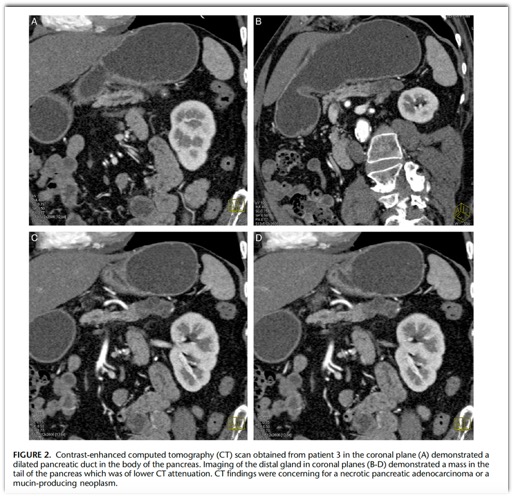
Japanese Journal of Radiology (2024) 42:246–260 - The radiologic finding of focal stenosis of the main pancreatic duct is highly suggestive of pancreatic cancer. Even in the absence of a mass lesion, focal duct stenosis can lead to surgical resection of the affected portion of the pancreas. We present four patients with distinctive pathology associated with non-neoplastic focal stenosis of the main pancreatic duct. The pathology included stenosis of the pancreatic duct accompanied by wavy, acellular, serpentine-like fibrosis, chronic inflammation with foreign body–type giant cell reaction, and calcifications. In all cases, the pancreas toward the tail of the gland had obstructive changes including acinar drop-out and interlobular and intralobular fibrosis. Three of the four patients had a remote history of major motor vehicle accidents associated with severe abdominal trauma. These results emphasize that blunt trauma can injure the pancreas and that this injury can result in long term complications, including focal stenosis of the main pancreatic duct. Pathologists should be aware of the distinct pathology associated with remote trauma and, when the pathology is present, should elicit the appropriate clinical history.
Distinctive Pathology Associated With Focal Stenosis of the Main Pancreatic Duct Secondary to Remote Trauma: A Long-term Complication of Seat Belt Pancreatitis.
Wu AA, Thompson ED, Cameron JL, He J, Burkhart RA, Burns WR, Lafaro KJ, Shubert CR, Canto MI, Fishman EK, Hruban RH.
Am J Surg Pathol. 2024 Mar 14. doi: 10.1097/PAS.0000000000002207. Epub ahead of print. PMID: 38482693. - The radiologic finding of focal stenosis of the main pancreatic duct is highly suggestive of pancreatic cancer. Even in the absence of a mass lesion, focal duct stenosis can lead to surgical resection of the affected portion of the pancreas. We present four patients with distinctive pathology associated with non-neoplastic focal stenosis of the main pancreatic duct. The pathology included stenosis of the pancreatic duct accompanied by wavy, acellular, serpentine-like fibrosis, chronic inflammation with foreign body–type giant cell reaction, and calcifications. In all cases, the pancreas toward the tail of the gland had obstructive changes including acinar drop-out and interlobular and intralobular fibrosis. Three of the four patients had a remote history of major motor vehicle accidents associated with severe abdominal trauma. These results emphasize that blunt trauma can injure the pancreas and that this injury can result in long term complications, including focal stenosis of the main pancreatic duct. Pathologists should be aware of the distinct pathology associated with remote trauma and, when the pathology is present, should elicit the appropriate clinical history.
Distinctive Pathology Associated With Focal Stenosis of the Main Pancreatic Duct Secondary to Remote Trauma : A Long-term Complication of Seat Belt Pancreatitis Annie A.
Wu, MD, PhD,* Elizabeth D. Thompson, MD, PhD,*† John L. Cameron, MD,‡ Jin He, MD, PhD,‡ Richard A. Burkhart, MD,‡ William R. Burns, MD,‡ Kelly J. Lafaro, MD, MPH,‡ Christopher R. Shubert, MD, MHA,‡ Marcia I. Canto, MD, MHS,†§ Elliot K. Fishman, MD,∥ and Ralph H. Hruban, MD*†
Am J Surg Pathol 2024;00:000–000) (in press) - “Four cases of non-neoplastic cutoff of the main pancreatic duct with distinctive pathology are presented. The pathology included abrupt duct stenosis, changes suggestive of rupture of the pancreatic duct, wavy, acellular, serpentine-like stromal fibrosis, and a foreign body– type giant cell reaction. IgG4 immunohistochemical stain was below the threshold in all cases, ruling out a type 1 autoimmune pancreatitis, and no neoplasms were present. Three of the four patients had a history of major motor vehicle accidents with associated severe abdominal trauma, suggesting that the unique pathology was caused by the trauma.”
Distinctive Pathology Associated With Focal Stenosis of the Main Pancreatic Duct Secondary to Remote Trauma : A Long-term Complication of Seat Belt Pancreatitis Annie A.
Wu, MD, PhD,* Elizabeth D. Thompson, MD, PhD,*† John L. Cameron, MD,‡ Jin He, MD, PhD,‡ Richard A. Burkhart, MD,‡ William R. Burns, MD,‡ Kelly J. Lafaro, MD, MPH,‡ Christopher R. Shubert, MD, MHA,‡ Marcia I. Canto, MD, MHS,†§ Elliot K. Fishman, MD,∥ and Ralph H. Hruban, MD*†
Am J Surg Pathol 2024;00:000–000) (in press) 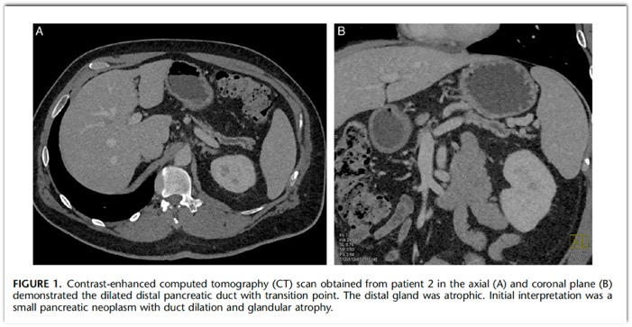


- “ In fact, the injured pancreas may appear normal on CT images, particularly in the first 12 hours after a trauma injury. If the admission CT image shows a normal pancreas but the patient subsequently develops abdominal pain, a repeat CT study obtained 24-48 hours later may show an injury not evident initially.”
Multidetector CT of Blunt Abdominal Trauma
Soto JA, Anderson SW
Radiology 2012; 265:678-693 - Pancreatic Trauma: Facts
- Neck and body are the most common sites of injury
- Pancreatic injuries can be classified as contusion, laceration or transection
- Injury to the pancreatic duct is associated with most severe injuries