Imaging Pearls ❯ Spleen ❯ Infection and Infarction
|
-- OR -- |
|
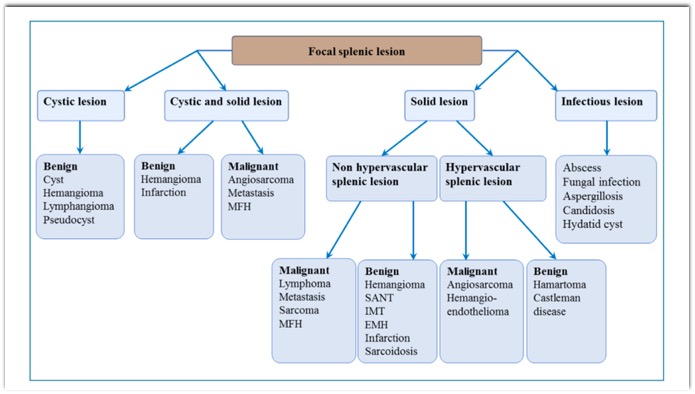
Diagnosis and treatment of focal splenic lesions
B. Malgras, H. Najah, A. Dohan et al.,
Journal of Visceral Surgery, https://doi.org/10.1016/j.jviscsurg.2021.11.010- “ Pyogenic Splenic abscess is most often due to hematogenous spread of infection with risk factors of diabetes, immunosuppression, corticosteroid therapy and sickle cell disease. It usually manifests as a high fever with chills and pain in the left hypochondrium. Splenic abscess can be single, multiple, or multiloculated.”
Diagnosis and treatment of focal splenic lesions
B. Malgras, H. Najah, A. Dohan et al.,
Journal of Visceral Surgery, https://doi.org/10.1016/j.jviscsurg.2021.11.010 - "On imaging, splenic candidiasis presents as multiple focal lesions that are small (< 1 cm) and rounded. They are hypoechoic on US, and minimally enhanced on CT and MRI. They are best visualized as hyper-intense on T2-weighted sequences and on diffusion sequences. Splenic candidiasis is most often associated with hepatic candidiasis, with hepatic lesions that show the same characteristics as the splenic lesions on imaging. MRI is suggestive when it reveals the ‘‘bull’s eye’’ sign (echogenic center sur- rounded by a hypo-echoic zone).”
Diagnosis and treatment of focal splenic lesions
B. Malgras, H. Najah, A. Dohan et al.,
Journal of Visceral Surgery, https://doi.org/10.1016/j.jviscsurg.2021.11.010 - “Sarcoidosis can affect all organs of the body; its manifesta- tions are non-specific, with clinical repercussions involving mainly the lungs, abdomen (pain) or systemic symptoms (fever, fatigue, weight loss). Imaging reveals splenic involve- ment in 6 to 33% of patients with sarcoidosis, although the prevalence of splenic involvement is between 24 and 59% when systematic histological analysis is performed, or even 38 to 77% in autopsy series. Splenic sarcoidosis is generally manifested by homogeneous splenomegaly that is present in approximately 40% of patients and much more rarely by focal splenic lesions.”
Diagnosis and treatment of focal splenic lesions
B. Malgras, H. Najah, A. Dohan et al.,
Journal of Visceral Surgery, https://doi.org/10.1016/j.jviscsurg.2021.11.010 - “When focal splenic lesions exist, they are often multi- ple, small in size (between 1 mm and 3 cm). They are rarely visible on US, and when they are, they are mildly hyperechoic. On CT, these lesions are mostly hypodense and contain small calcifications in 16% of cases. After IV contrast injection, the lesions are hypodense compared to the splenic parenchyma without peripheral enhancement. On MRI, the focal splenic lesions of sarcoidosis are hypo-intense with weak and late enhancement. They are better visible in T2-weighting with fat saturation, or in the early phase after injection. The lesional architecture is similar to that of lymphomas or splenic metastases.”
Diagnosis and treatment of focal splenic lesions
B. Malgras, H. Najah, A. Dohan et al.,
Journal of Visceral Surgery, https://doi.org/10.1016/j.jviscsurg.2021.11.010