Imaging Pearls ❯ Colon ❯ Volvulus
|
-- OR -- |
|
- “Cecal volvulus is torsion of the cecum and ascending colon, accounting for 1–3% of cases of large-bowel obstruction. The progression of cecal volvulus can result in bowel ischemia, necrosis, or perforation, as well as death. Rapid surgical treatment can treat, or even prevent, these complications. Abdominal radiography is suboptimal for the diagnosis, having a re- ported sensitivity of 75%, and being diagnostic in only 17% of patients.”
Utility of CT Findings in the Diagnosis of Cecal Volvulus Dane B et al. AJR 2017; 209:762–766 - “The readers also assessed images for a proposed new potential finding of cecal volvulus that we designate as the “central appendix” sign. The central appendix sign was defined as an abnormal position of the appendix near mid- line, whereby the appendix is rotated toward the right side of the cecum and is on the same side as the terminal ileum.”
Utility of CT Findings in the Diagnosis of Cecal Volvulus Dane B et al. AJR 2017; 209:762–766 - “The radiologists evaluated the CT examinations for the presence or absence of the following features; whirl sign (previously defined as collapsed loops of cecum with swirling strands of soft tissue, vessels, and fat attenuation centrally, abnormal cecal position (defined as cecum located outside the right lower quadrant), “bird beak” sign (previously defined as progressive tapering of afferent and efferent loops at the site of torsion), severe cecal distention, and mesenteric engorgement.”
Utility of CT Findings in the Diagnosis of Cecal Volvulus Dane B et al. AJR 2017; 209:762–766 - “CT exhibited excellent diagnostic performance and very high sensitivity for cecal volvulus for readers of varying experience. Secondary imaging ndings may be useful in interpretation given that oral contrast material often fails to reach the transition point in patients with this
diagnosis. The previously described whirl sign was a signi cant independent predictor of cecal volvulus for both readers. The newly proposed central appendix sign may also have potential value for less experienced readers.”
Utility of CT Findings in the Diagnosis of Cecal Volvulus Dane B et al. AJR 2017; 209:762–766
- Cecal Volvulus
- Cecal Volvulus
- “Volvulus of the cecum is a torsion of the bowel around its own mesentery that often results in a closed- loop obstruction. Cecal volvulus can only occur in the small percentage (11–25%) of the population who have a developmental failure of peritoneal fixation, allowing the proximal colon to be free and mobile. The second requirement is restriction of the bowel at a fixed point within the abdomen that serves as a fulcrum for rotation, such as an adhesion, abdominal mass, or scarring
from calcified lymph nodes.“
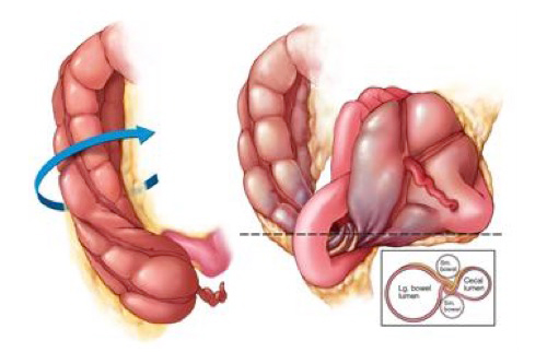
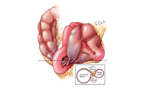
CT of Cecal Volvulus: Unraveling the Image Carolyn J. Moore, Frank M. Corl, Elliot K. Fishman AJR Am J Roentgenol. 2001 Jul;177(1):95-8 - “Cecal volvulus accounts for 11% of all in- testinal volvulus, generally occurring in pa- tients who are 30–60 years old. Medical history of these patients may include prior abdominal surgery, presence of a pelvic mass, violent coughing, atonia of the colon, extreme exertion, unpressurized air travel, or third-trimester pregnancy. Patients present with nausea, vomiting, constipation, and acute cramping pain.” CT of Cecal Volvulus: Unraveling the Image Carolyn J. Moore, Frank M. Corl, Elliot K. Fishman AJR Am J Roentgenol. 2001 Jul;177(1):95-8
- “Three-dimensional imaging is ideal because, like a barium enema, the entire bowel can be visualized in a single image, separating the volvulus from other dilated loops. Three-dimensional displays allow radiologists to select the optimal plane for viewing the volvulus and to locate the precise
source of the torsion . In effect, the ability to analyze an image in multiple planes allows one to unravel twisted bowel and confirm the diagnosis of volvulus. .”
CT of Cecal Volvulus: Unraveling the Image Carolyn J. Moore, Frank M. Corl, Elliot K. Fishman AJR Am J Roentgenol. 2001 Jul;177(1):95-8 - “On axial CT images, cecal volvulus is sug- gested by the extreme dilatation of the cecum. When seen on conventional radiographs or to- mograms, the cecal volvulus is seen as a rounded focal collection of air-distended bowel with haustral creases in the left upper quadrant that resembles a coffee bean . The two limbs of the looped obstruction gradually taper and converge at the site of the torsion, resulting in the appearance of a bird’s bea.”
CT of Cecal Volvulus: Unraveling the Image Carolyn J. Moore, Frank M. Corl, Elliot K. Fishman AJR Am J Roentgenol. 2001 Jul;177(1):95-8 - “The reduction rates in cecal volvulus achieved through use of colonoscopy are much lower than those achieved in sigmoid volvulus, and in patients with cecal volvulus, the recurrence rate exceeds 50%. In patients with uncomplicated cecal volvulus, surgical options include cecopexy, which has a low rate of morbidity (0–8%) and volvulus recurrence.”
CT of Cecal Volvulus: Unraveling the Image Carolyn J. Moore, Frank M. Corl, Elliot K. Fishman AJR Am J Roentgenol. 2001 Jul;177(1):95-8


"Two defined transition points are seen in the minority of cases of cecal volvulus; a single transition point is more sensitive for cecal volvulus."
Findings of Cecal Volvulus at CT
Rosenblat JM et al.
Radiology 2010; 256;169-175"When cecal volvulus is suspected, the absence of distal colonic decompression on CT topograms makes the diagnosis very unlikely. Whirl, ileocecal twist, transition points, X-marks-the-spot, and split wall have high specificity for cecal volvulus."
Findings of Cecal Volvulus at CT
Rosenblat JM et al.
Radiology 2010; 256;169-175