Imaging Pearls ❯ Chest ❯ Cinematic Rendering
|
-- OR -- |
|
- “Routinely employed 3D post-processing tools include maximum intensity projection (MIP) and volume rendering (VR) that allow for the generation of angiographic images and a more intuitive and interactive representations of the spatial information in the dataset, respectively. Improving upon traditional VR, 3D cinematic rendering (CR) is a Food and Drug Administration (FDA)-approved technique that employs a novel lighting model to generate photorealistic images. CR involves global illumination and path tracing models whereby numerous light rays from all directions propagate through and interact with the volumetric data to generate a voxel. Complex anatomical relations are better evaluated and enhanced depth and shape perception is achieved as the technique considers a natural lighting environment and its effects (e.g., reflection, diffusion, refraction). Postprocessing windowing and the use of clip planes/masks allow cutting into the volume and isolation of the area/organ of interest.”
Cinematic rendering of non-traumatic thoracic aorta emergencies: a new look at an old problem.
Yasrab M, Rizk RC, Chu LC, Fishman EK.
Emerg Radiol. 2024 Jan 18. doi: 10.1007/s10140-024-02204-6. Epub ahead of print. - “There are some limitations that come with 3D CR. Notably, shadows generated in the images might conceal certain pathologies when viewed from specific angles, necessitating meticulous optimization and assessment from diverse angles in conjunction with the multiplanar reformations. Thus, while an initial learning period to become adept in handling and familiarizing themselves with the CR process is required for radiologists, as demonstrated in our case studies, an experienced radiologist can efficiently execute the rendering process in under 5 min.”
Cinematic rendering of non-traumatic thoracic aorta emergencies: a new look at an old problem.
Yasrab M, Rizk RC, Chu LC, Fishman EK.
Emerg Radiol. 2024 Jan 18. doi: 10.1007/s10140-024-02204-6. Epub ahead of print. - “Color mapping of different phases enhances visualization of the key pathology, such as the flow through the false and true lumens in a dissection that can be delineated by high contrast shading. CR rendering emphasizes textural changes attributable to inflammatory processes with realistic shadowing that is otherwise difficult to appreciate. The improved surface detail helps characterize an impending PAU or the nature of outpouchings suspicious for mycotic aneurysms and gives a clearer view of multiple plaques and sites of ulceration that could be otherwise missed.”
Cinematic rendering of non-traumatic thoracic aorta emergencies: a new look at an old problem.
Yasrab M, Rizk RC, Chu LC, Fishman EK.
Emerg Radiol. 2024 Jan 18. doi: 10.1007/s10140-024-02204-6. Epub ahead of print. - “Another application of 3D CR is via the black blood cinematic rendering (BBCR) preset.. BBCR is a preset we specifically developed to visualize intraluminal contours and structures of the heart and great vessels, all through adjustments that can be madein under a minute. This is especially useful in the setting of visualizing various zones of thrombi and occlusion, the degree of obstruction, and the subtle irregularities and internal arrangement of the thrombus that can only be appreciateddue to enhanced depth perception and shadowing.”
Cinematic rendering of non-traumatic thoracic aorta emergencies: a new look at an old problem.
Yasrab M, Rizk RC, Chu LC, Fishman EK.
Emerg Radiol. 2024 Jan 18. doi: 10.1007/s10140-024-02204-6. Epub ahead of print. - “The intrinsic features of 3D CR with its ability to provide a holistic field of view of the vascular map increases confidence in management and surgical planning. A global viewing angle of the thoracic aorta helps in tracking the dissection and its involvement of the aorta and the extent of mediastinal and pericardial bleeding where present .In cases where patients underwent thoracic endovascular aortic repair (TEVAR), coiling, or graft repairing, CR adds to surgical planning by improved depth perception, shadow effects, and realistic textures, demonstrating the anatomical relationships of the thoracic aorta, surrounding structures, and the pathology to be addressed, with photorealism providing the surgeon a familiar perspective to work with.”
Cinematic rendering of non-traumatic thoracic aorta emergencies: a new look at an old problem.
Yasrab M, Rizk RC, Chu LC, Fishman EK.
Emerg Radiol. 2024 Jan 18. doi: 10.1007/s10140-024-02204-6. Epub ahead of print. - “3D cinematic rendering (CR) represents an important advancement in radiological imaging, particularly in enhancing the visualization of complex anatomical structures and systems such as the thoracic aorta and its vessels. CR provides detailed, photorealistic illustrations crucial for diagnosis and surgical planning as we have seen in several cases. Future research is needed to evaluate CR’s diagnostic accuracy, both prospectively and in head-to-head comparisons with other rendering methods, as well as its role in other domains such as patient education and medical training. CR, therefore, is emerging as a promising, evolving tool for radiologists, surgeons, and the patients they treat.”
Cinematic rendering of non-traumatic thoracic aorta emergencies: a new look at an old problem.
Yasrab M, Rizk RC, Chu LC, Fishman EK.
Emerg Radiol. 2024 Jan 18. doi: 10.1007/s10140-024-02204-6. Epub ahead of print.
- ANCA Vasculitis
Antineutrophilic cytoplasmic antibody (ANCA) associated vasculitides are a heterogeneous group of rare autoimmune conditions that causes an inflammation of blood vessels with various manifestations. It includes three main diseases, which are granulomatosis with polyangiitis (GPA; formerly known as Wegener granulomatosis), eosinophilic granulomatosis with polyangiitis (EGPA; previously known as Churg-Strauss syndrome), and microscopic polyangiitis (MPA). Other ANCA-associated diseases are drug-induced vasculitis and renal limited vasculitis.. - ANCA Vasculitis
Antineutrophilic cytoplasmic antibody (ANCA) associated vasculitides are rare diseases. The incidence reported being 10 to 20 cases per million. GPA is the most common disease of the three types, with an incidence of 5 to 10 cases per million and a peak incidence in middle age (approximately 55 years). MPA is less common than GPA with a male to female ratio of 2:1, and EGPA is the rarest of all. 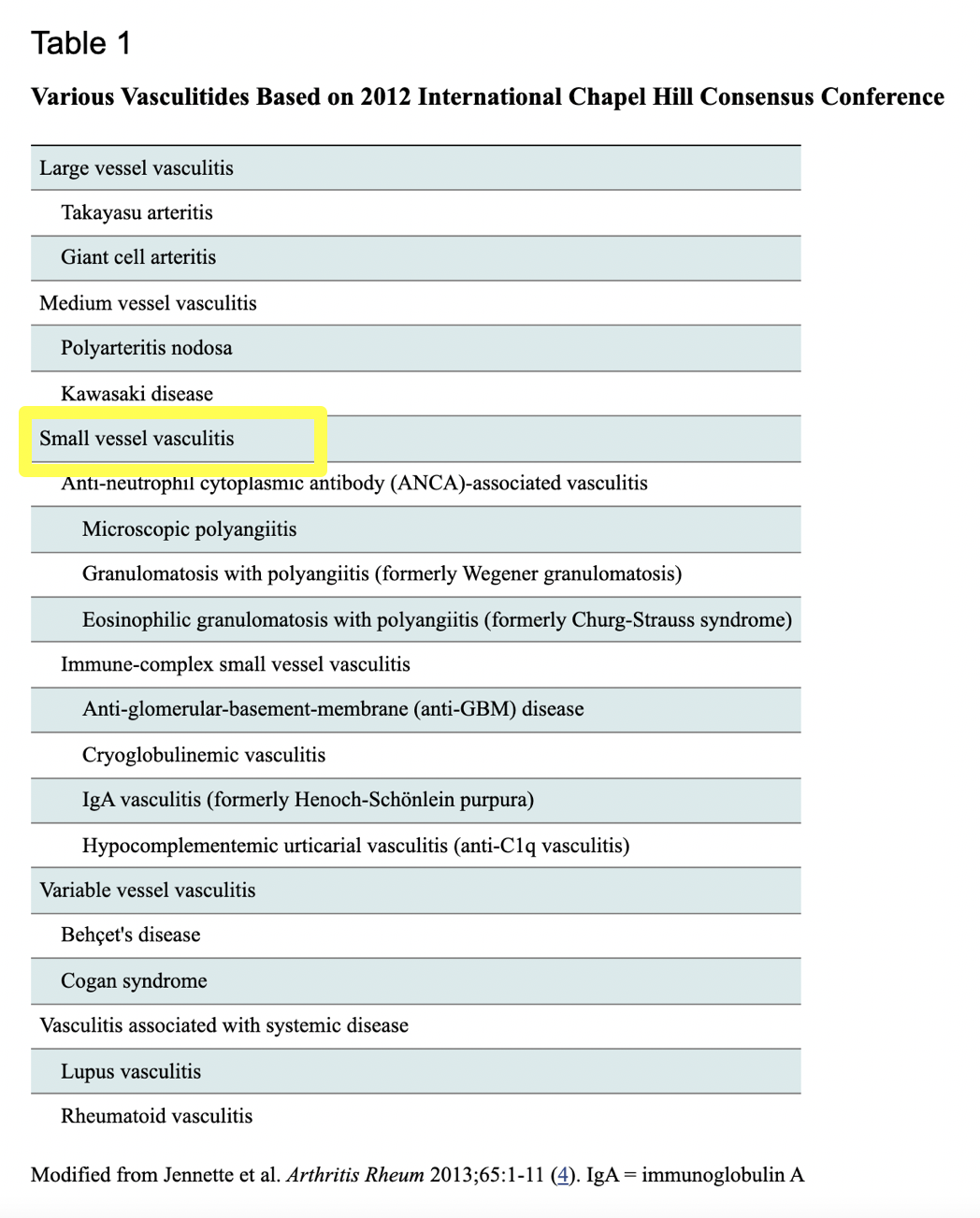
- ANCA Vasculitis
In GPA, pulmonary nodules or masses are the most common findings, which are usually multiple and bilateral. Central cavitation may be noted in up to 50% of nodules larger than 2 cm in diameter. Areas of consolidation or ground-glass opacity (GGO) are also noted in 30% of patients with active GPA, which may be the result of alveolar hemorrhage or mosaic perfusion secondary to small vessel vasculitis.
- “Utilizing complex lighting models, cinematic rendering is a novel technique for demonstrating computed tomography data with exquisite 3D anatomic detail. The tracheal lumen, tracheal wall, and adjacent soft tissue structures are represented with photorealistic detail exceeding that of conventional volume rendering or virtual bronchoscopy techniques. We applied cinematic rendering to a spectrum of emergent tracheal pathologies: traumatic tracheal tears, tracheoesophageal fistulas, tracheal foreign bodies, tracheal stenosis (intrinsic and extrinsic causes), tracheal neoplasms, and tracheomalacia. Cinematic rendering images enable visually accessible evaluation and comprehensive understanding of acute tracheal pathology, which is likely to be of value to both interventional pulmonologists and thoracic surgeons who are determining patient treatment plans.”
Cinematic rendering enhancements to virtual bronchoscopy: assessment of emergent tracheal pathology
Cheng Ting Lin, Steven Rowe, Linda C. Chu, Hannah Recht, Elliot K. Fishman
Emergency Radiology (2021) 28:193–199 - "Cinematic rendering (CR) is a novel technique for demonstrating the same data with 3D anatomic detail via the use of complex lighting models. This method fully visualizes the relationship of the trachea to adjacent mediastinal structures with a photorealistic level of detail that is not available with conventional volume rendering or virtual bronchoscopy techniques. CR utilizes a more complex lighting model that more accurately depicts the manner in which photons act when coming into contact with real-world objects.”
Cinematic rendering enhancements to virtual bronchoscopy: assessment of emergent tracheal pathology
Cheng Ting Lin, Steven Rowe, Linda C. Chu, Hannah Recht, Elliot K. Fishman
Emergency Radiology (2021) 28:193–199 - "Tracheal stenosis is fixed airway narrowing caused by various congenital and acquired disorders. The severity of stenosis generally stays stable during respiration, although occasionally, tracheomalacia may coexist. Severe and extensive tracheal stenosis obstructs airflow and requires urgent intervention. Via multiplanar reformats and 3D rendering, CT imaging pro- vides reliable diagnostic information that aids in preprocedural planning and subsequent posttreatment surveillance.”
Cinematic rendering enhancements to virtual bronchoscopy: assessment of emergent tracheal pathology
Cheng Ting Lin, Steven Rowe, Linda C. Chu, Hannah Recht, Elliot K. Fishman
Emergency Radiology (2021) 28:193–199 - “Tracheomalacia is defined by excessive weakness and collapsibility of the airway, leading to transient expirato- ry collapse. Traditionally, the diagnosis is made with bronchoscopy by directly visualizing airway collapse during forced exhalation. A dynamic decrease in luminal cross-sectional area of at least 50% compared to end- inspiration is considered diagnostic, although this threshold can be exceeded by some healthy volunteers; therefore, 70% collapse would be a more specific criterion. Using a 3D technique is valuable for global visualization of the tracheobronchial tree and the extent of airway collapse beyond that visible on bronchoscopy.”
Cinematic rendering enhancements to virtual bronchoscopy: assessment of emergent tracheal pathology
Cheng Ting Lin, Steven Rowe, Linda C. Chu, Hannah Recht, Elliot K. Fishman
Emergency Radiology (2021) 28:193–199 - "CR images enable visually accessible evaluation and comprehensive understanding of acute tracheal pathology. The tracheal lumen, tracheal wall, and adjacent soft tissue structures are represented with photorealistic detail exceeding that of conventional volume rendering. Assessment of tracheal disorders using CR confers several advantages, including clear delineation of the spatial relationship between the trachea and surrounding structures, rapid recognition of the configuration and severity of tracheal stenosis, and improved visualization distal to the occluded airway.”
Cinematic rendering enhancements to virtual bronchoscopy: assessment of emergent tracheal pathology
Cheng Ting Lin, Steven Rowe, Linda C. Chu, Hannah Recht, Elliot K. Fishman
Emergency Radiology (2021) 28:193–199