- Atrial septal aneurysm
- Bulging of the fossa ovalis 10-15 mm beyond the tissue of interatrial septum into left or right atrium
- Associated with PFO in the majority of cases
- If filled with unopacified blood, can mimic an intracardiac mass - “An atrial septal aneurysm (ASA) is a rare but well recognized and localized saccular deformity of the atrial septum that bulges into the right or left atrium with uncertain clinical significance. Diagnosis can be established using transthoracic (TTE) and transesophageal echocardiography. Although these abnormalities are considered clinically benign entities, they have been independently associated with ischemic stroke.”
Atrial septal aneurysm and stroke
Mohd Razaq R et al.
Ann Pediatr Cardiol. 2012 Jan-Jun; 5(1): 98–99. - “ASA is an uncommon congenital car- diac abnormality, characterized by a diffuse or localized protrusion of the interatrial septum into the right or left atrium, or both. The prevalence of ASA is 0.2%–3% in the general population. The MDCT features of this anomaly were described for the first time, in 2006, by Zeina et al.”
Usefulness of 64-Slice Computed tomography for evaluation of atrial Septal aneurysm
Zeina AR
IMAJ 2011; 13: 645–646 - “An AMS is an uncommon congenital defect frequently found in conjunction with ventricular septal defects; discovery might be incidental, as it was for this patient. These lesions are usually grouped with other defects of the left ventricular outflow tract and have been diagnosed by means of angiography, echocardiography, and magnetic resonance imaging. However, reports of the discovery of this defect on CCT are uncommon.”
Membranous Ventricular Septal Aneurysm
Diagnosed by Means of Cardiac Computed Tomography
Afaneh AB et al.
Texas Heart Institute Journal
Volume 39, Number 3, 2012 - “Although most cases do not manifest themselves symptomatically, AMS can be associated with systemic emboli, endocarditis, cardiac arrhythmias, left or right ventricular outflow tract obstruction, and right-to-left shunts secondary to ruptures.”
Membranous Ventricular Septal Aneurysm
Diagnosed by Means of Cardiac Computed Tomography
Afaneh AB et al.
Texas Heart Institute Journal
Volume 39, Number 3, 2012 - “For this patient, CCT and volume imaging acquisition provided a diagnostic advantage by reason of its multiplanar 3-dimensional reconstructions. The increasing availability of multiplanar cardiac imaging techniques has improved the accuracy of diagnosis for many cardiac diseases. Magnetic resonance imaging and CCT have important roles in the evaluation of cardiac aneurysms.”
Membranous Ventricular Septal Aneurysm
Diagnosed by Means of Cardiac Computed Tomography
Afaneh AB et al.
Texas Heart Institute Journal
Volume 39, Number 3, 2012
- “Interatrial septal aneurysm (IASA) consists of redundant atrial septal tissue, which bulges into either the left or the right atrium. The clinical implications of this entity are not entirely clear; however, if it is associated with other cardiac abnormalities such as patent foramen ovale and atrial septal defects. It may assume significance by increasing the risk of cardioembolic events such as stroke. We present a case of an individual with giant IASA detected by transesophageal echocardiography, which was mimicking a left atrial mass on transthoracic echocardiography.”
Giant interatrial septal aneurysm mimicking a left atrial mass.
Taleb M et al.
Heart Views. 2013 Apr;14(2):88-9 - “IASA is a congenital malformation of the atrial septum that may occur as an isolated abnormality or in association with various cardiac defects, such as patent foramen ovale (PFO), atrial septal defect (ASD), and mitral valve prolapse or connective tissue diseases. [1],[2] Its prevalence varies depending on the diagnostic method used but with the widespread use of echocardiography, IASA has become an increasingly recognized entity.”
Giant interatrial septal aneurysm mimicking a left atrial mass.
Taleb M et al.
Heart Views. 2013 Apr;14(2):88-9 - “ The diagnosis of IASA is very important to prevent various complications and in differentiating it from an intracardiac mass-like myxoma which will have implications on choice of therapy.”
Giant interatrial septal aneurysm mimicking a left atrial mass.
Taleb M et al.
Heart Views. 2013 Apr;14(2):88-9 - “The Amplatzer septal occluder is very efficient and offered interventional ASD closure in 84.7% of our group of consecutive patients, with excellent intermediate results.”
Experience with transcatheter closure of secundum atrial septal defects using the Amplatzer septal occluder: a single centre study in 236 consecutive patients
G Fischer et al.
Heart Feb 2003; 89(2):199-204 - “The ease of implantation and the superior success rate of ASD closure with the Amplatzer septal occluder has led to the widespread employment of transcatheter occlusion of ASD and has replaced routine surgical closure in many centres. There is no doubt that this success rate is a result of the design of the Amplatzer occluder, which is completely different from the patch type systems. The most important aspect is that the device’s waist between the left and right retention discs is a stent, resulting in self centring within the defect.”
Experience with transcatheter closure of secundum atrial septal defects using the Amplatzer septal occluder: a single centre study in 236 consecutive patients
G Fischer et al.
Heart Feb 2003; 89(2):199-204 - “The AMPLATZER Septal Occluder is a device specifically designed to close an ASD. The device is implanted during a catheter-based procedure and remains permanently implanted. The device is made from a braided metal (Nitinol) that has shape memory characteristics; this means the device will go back to its original shape even after it is stretched to pass through a catheter.”
St. Jude Medical 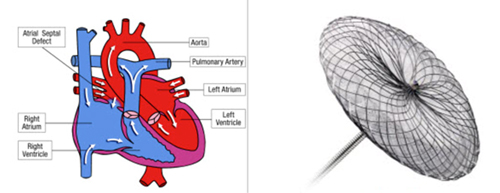
- Atrial Myxomas: Facts
- Most common benign neoplasm of the heart
- Accounts for 50% of benign cardiac tumors
- Mean age of diagnosis is 50 years of age
- Patient presentation ranges from arrhythmias, symptoms of intracardiac obstruction , systemic embolization and/or constitutional symptoms
- Vast majority attach to the fossa ovalis of the interatrial septum
- 75% in left atrium and 25% in right atrium - “ Although cardiac masses are rare entities, patients with cardiac masses may present with acute symptoms and may be encountered by emergency radiologists. Detection and tailored differential diagnosis of cardiac masses are important in guiding management.”
Multidetector CT of the heart: spectrum of benign and malignant cardiac tumors
Chu LC, Johnson PT, Halushka MK, Fishman EE
Emerg Radiol (2012) 19:415-428 - “ In general we recommend screening with TTE followed by TEE in patients with clinical heart valve dysfunction. When results of TTE and TEE are inconclusive, use of cardiac CT and MR imaging should be considered. If there are contraindications to CT, then MR imaging should be used and visa versa.”
Complications of Aortic Valve Surgery: Manifestations at CT and MR Imaging
Pham N et al.
RadioGraphics 2012; 32:1873-1892
- Cardiac Masses in the Interatrial Septal Region
-Atrial myxomas
-Metastatic tumors with common locations of origin include;
-Lung cancer
-Breast cancer
-Melanoma (highest percent)
-Lymphoma - Lipomatous hypertrophy of the interatrial septum: Facts
-Defined as an accumulation in the IAS of adipose tissue exceeding 15 mm and which spares the fossa ovalis
-Is seen by CT and echo in just under 3% of patients
-Occurs more commonly in elderly and obese patients and associated with abnormalities in venous drainage, atrial arrhythmias (40% of patients) and sudden cardiac death
-Classic dumbbell appearance on CT - Cor triatriatum: facts
-0.1% of congenital heart disease
-Consists of fibromuscular band that divides the left atrium into 2 chambers
-Usually a flap extends from the interatrial septum and extending to the atrial free wall
-Associated with ASD, mitral regurgitation, coronary sinus malformations
-Usually presents in infancy but may be seen in adults presenting with symptoms due to long standing pulmonary venous congestion (dyspnea, orthopnea, hemoptysis) - Atrial septal aneurysms: facts
-Atrial septal aneurysms is defined as an abnormal protrusion of the flap valve with an excursion of ? 10 mm beyond the plane of the IAS with a base of ? 15mm
-Autopsy prevalence of 1%
-May be an isolated finding or seen with patent foramen ovale, AD and sinus venosus defect
-Can mimic myxoma on non-gated images - Interatrial Septal Pathology
-Atrial septal aneurysms
-Lipomatous hypertrophy of the interatrial septum
-Cor triatriatum
-Masses (atrial myxoma, metastatic tumors)
-Thrombus - “ Interpretation of the complex structures of the interatrial region requires a detailed knowledge of septal anatomy, variants, and atrial septal disorders.”
Cardiac CT of non-shunt pathology of the interatrial septum
Rojas CA et al.
J Cardiovascular Comput Tomogr (2011) 5, 93-100 - CT Features that distinguish benign and malignant tumors
Feature
Benign
Malignant
location
More on left side
More in right atrium
margin
Smooth, well defined
Lobulated, ill defined, invasive,infiltrative
invasion
none
Myocardium,pericardium,extracardiac attachment
Pedicle may be seen
Broad based
Feeding vessel
absent
May be present
calcification
Rare,except in myxoma
Large foci in osteosarcoma
Pericardial effusion
Not seen
Suspicious for malignancy
metastasis
none
May be present
- Cardiac Myxoma: Clinical Presentation
-Valvular or intracavitary dysfunction
-Embolism (seen in 35% of left sided myxomas)
-Constitutional symptoms like fever, malaise, and weight loss - Cardiac Myxoma: CT Findings
-Well defined, lobulated, smooth or oval mass
-Calcification common
-Patchy enhancement on contrast enhanced CT
-May prolapse through cardiac valves
-Most common in left atrium - Left Atrial Appendage Thrombi Protocol
- Scan 1: trigger the injection 6 seconds after 100 HU is reached in ascending aorta
- Scan 2: this scan is 30 seconds after first scan is completed
- Injection protocol was 60-90 cc of Iopamidol-370 injected at 5 cc/sec "Two phase cardiac CT angiography can be used to differentiate thrombus from circulatory stasis, which may cause a pseudo-filling defect on early phase CT images."
Left Atrial Appendage Thrombi in Stroke Patients: Detection with Two-Phase Cardiac CT Angiography versus Transesophageal Echocardiography
Hur J et al.
Radiology 2009; 251:683-690"Compared with transesophageal echocardiography (TEE), two phase cardiac CT angiography, with a sensitivity of 100% and a specificity of 98%, is useful for detecting left atrial appendage thrombus."
Left Atrial Appendage Thrombi in Stroke Patients: Detection with Two-Phase Cardiac CT Angiography versus Transesophageal Echocardiography
Hur J et al.
Radiology 2009; 251:683-690"Two phase 64-section cardiac CT angiography is a noninvasive sensitive modality for detecting left atrial appendage thrombi and differentiating thrombus from circulatory stasis in stroke patients."
Left Atrial Appendage Thrombi in Stroke Patients: Detection with Two-Phase Cardiac CT Angiography versus Transesophageal Echocardiography
Hur J et al.
Radiology 2009; 251:683-690"Our data show that left atrial accessory appendages and diverticula can be found in more than one fifth of subjects undergoing cardiac CTA and are more common in men.Accessory left atrial appendages tend to be smaller than diverticula and are more commonly found on the left lateral atrial wall. Whether there is any pathologic value remains uncertain."
Cardiac CT Assessment of Left Atrial Accessory Appendages and Diverticula
Abbara S et al.
AJR 2009; 193:807-812"Cor triatriatum is a rare congenital heart disease (CHD) estimated to occur in 0.1% to 0.5% of patients with CHD. In cor triatriatum a fibromuscular membrane divides the left atrium into a posterosuperior proximal cavity and an anteroinferior distal cavity."
Asymptomatic Cor Triatriatum: Utility of 64-Slice Multidetector Computed Tomography with 3-Dimensional Volume Rendering
Shan SJ, Johnson PT, Fishman EK
J Comput Assist Tomogr 2009;33: 779-781- Cardiac Myxomas: Facts
- Most common benign primary tumor of the heart
- Arises from left atrium near interatrial septum
- Most difficult dx in many cases is an cardiac thrombus - Atrial Myxoma vs Thrombus: Key Differential Dx Parameters
- CT attenuation of mass
- Size of mass
- Left vs right atrium
- Origin of mass
- Lesion shape
- Lesion mobility
- Occurrence of prolapse "Atrial myxoma and thrombi can be differentiated by their distinguishing features of size, origin, shape, mobility and prolapse. CT is accurate in determining the origin of myxomas but may fail in some cases."
Atrial Myxomas and Thrombi: Comparison of Imaging Features on CT
Scheffel H et al.
AJR 2009; 192:639-645"The sensitivity of CT imaging is not sufficient for diagnosing patent foramen ovale. However if this characteristic finding is detected at routine cardiac CT, the presence of a patent foramen is strongly suggested."
Patent Foramen Ovale: Diagnosis with Multidetector CT-Comparison with Transesophageal EchocardiographyKim YJ et al.Radiology 2009; 250:61-67"A contrast agent jet from left atrium to right atrium toward the inferior vena cava with channel-like appearance of the intraatrial septum on CT images confirms the presence of a patent foramen ovale."
Patent Foramen Ovale: Diagnosis with Multidetector CT-Comparison with Transesophageal EchocardiographyKim YJ et al.Radiology 2009; 250:61-67- Atrial Myxoma: Facts
- Most common benign tumor of the heart
- 85% are in left atrium attached to atrial septum near fossa ovalis
- Calcification is seen in half the patients - Atrial Myxoma: Differential Diagnosis
- Intracardiac thrombus
- Cardiac metastasis
- Cardiac lipoma
- Primary cardiac malignancies
- Cardiac lymphoma