Imaging Pearls ❯ Cardiac ❯ Automated Analysis of the Coronary Arteries
|
-- OR -- |
|
- “Singh and colleagues provided an overview of artificial intelligence and machine learning that are pertinent to contemporary applications within cardiovascular imaging. They high- lighted areas within healthcare where machine learning could stream- line processes, as well as minimize inefficiencies. In addition, the application of neural networks for image segmentation was reviewed both in non-contrast and contrast CCT studies.”
The Journal of Cardiovascular Computed Tomography year in review - 2018
Al’Aref SJ et al.
Journal of Cardiovascular Computed Tomography (in press) 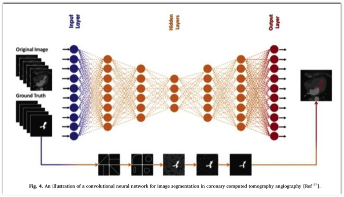
The Journal of Cardiovascular Computed Tomography year in review - 2018
Al’Aref SJ et al.
Journal of Cardiovascular Computed Tomography (in press)

- “ Structured reporting of incidental CT findings can mediate accurate stratification of individuals into clinically relevant risk categories and subsequently allow those at higher risk of future CVD events to be distinguished.”
Incidental Imaging Findings from Routine Chest CT Used to Identify Subjects at High Risk of Future Cardiovascular Events
Jairam PM et al.
Radiology 2014; 272:700-708 - “ The final model included patient age and sex, CT indication, left anterior descending coronary artery calcifications, mitral valve calcifications, descending aorta calcifications and cardiac diameter. This imaging based model allows accurate stratification of individuals into clinically relevant risk categories.”
Incidental Imaging Findings from Routine Chest CT Used to Identify Subjects at High Risk of Future Cardiovascular Events
Jairam PM et al.
Radiology 2014; 272:700-708
- “Automated software is a time-saving method that allows accurate assessment of coronary atherosclerotic plaque volumes in coronary CTA with high reproducibility. With this approach, serial studies appear to be possible.”
Interscan reproducibility of quantitative coronary plaque volume and composition from CT coronary angiography using an automated method.
Schuhbaeck A, Dey D, Otaki Y, Slomka P, Kral BG, Achenbach S, Berman DS, Fishman EK, Lai S, Lai H.Eur Radiol. 2014 Sep;24(9):2300-8. doi: 10.1007/s00330-014-3253-3. Epub 2014 Jun 25. - “Quantitative measurements of coronary plaque volume may play a role in serial studies to determine disease progression or regression. Our aim was to evaluate the interscan reproducibility of quantitative measurements of coronary plaque volumes using a standardized automated method.”
Interscan reproducibility of quantitative coronary plaque volume and composition from CT coronary angiography using an automated method.
Schuhbaeck A, Dey D, Otaki Y, Slomka P, Kral BG, Achenbach S, Berman DS, Fishman EK, Lai S, Lai H.Eur Radiol. 2014 Sep;24(9):2300-8. doi: 10.1007/s00330-014-3253-3. Epub 2014 Jun 25. - - Reproducibility of coronary atherosclerotic plaque volume in coronary CTA is high.
- Using automated software facilitates quantitative measurements.
- Serial studies to determine progression or regression of coronary plaque are possible”
Interscan reproducibility of quantitative coronary plaque volume and composition from CT coronary angiography using an automated method.
Schuhbaeck A, Dey D, Otaki Y, Slomka P, Kral BG, Achenbach S, Berman DS, Fishman EK, Lai S, Lai H.Eur Radiol. 2014 Sep;24(9):2300-8. doi: 10.1007/s00330-014-3253-3. Epub 2014 Jun 25.
- “Reproducibility of coronary atherosclerotic plaque volume in coronary CTA is high. Using automated software facilitates quantitative measurements. Serial studies to determine progression or regression of coronary plaque are possible.”
Interscan reproducibility of quantitative coronary plaque volume and composition from CT coronary angiography using an automated method.
Schuhbaeck A, Dey D, Otaki Y, Slomka P, Kral BG, Achenbach S, Berman DS, Fishman EK, Lai S, Lai H.
Eur Radiol. 2014 Jun 25. [Epub ahead of print] - “Coronary dual source computed tomography angiography (CTA) was performed twice in 20 consecutive patients with known coronary artery disease within a maximum time difference of 100 days. The total plaque volume (TP), the volume of non-calcified plaque (NCP) and calcified plaque (CP) as well as the maximal remodelling index (RI) were determined using automated software.”
Interscan reproducibility of quantitative coronary plaque volume and composition from CT coronary angiography using an automated method.
Schuhbaeck A, Dey D, Otaki Y, Slomka P, Kral BG, Achenbach S, Berman DS, Fishman EK, Lai S, Lai H.
Eur Radiol. 2014 Jun 25. [Epub ahead of print] - “Mean TP volume was 382.3?±?236.9 mm3 for the first and 399.0?±?247.3 mm3 for the second examination (p?=?0.47). There were also no significant differences for NCP volumes, CP volumes or RI. Interscan correlation of the plaque volumes was very good (Pearson's correlation coefficients: r?=?0.92, r?=?0.90 and r?=?0.96 for TP, NCP and CP volumes, respectively).”
Interscan reproducibility of quantitative coronary plaque volume and composition from CT coronary angiography using an automated method.
Schuhbaeck A, Dey D, Otaki Y, Slomka P, Kral BG, Achenbach S, Berman DS, Fishman EK, Lai S, Lai H.
Eur Radiol. 2014 Jun 25. [Epub ahead of print]
- “ Thus, there could be
displays with distinct properties that are explicitly four-dimensional (e.g., spatial–temporal, spatial–spectral) or five-dimensional (e.g., spatial–temporal–spectral) and be unique for a disease process.”
Adding Dimensions to Unimodal Cardiac Images
Sengupta PP, Marwick TH, Narula J
J Am Coll Cardiol Img 2011;4;816-818 - “The combination of functional and
morphological information processed through a single modality may have relevance for improving the understanding of complex biologic mechanisms involved in routine clinical imaging.”
Adding Dimensions to Unimodal Cardiac Images
Sengupta PP, Marwick TH, Narula J
J Am Coll Cardiol Img 2011;4;816-818 - “ Following further validation, the automated tool in this study could potentially be applied routinely to provide rapid noninvasive quantitative assessment of coronary plaques with coronary CT angiography.”
Automated Three-dimensional Quantification of Noncalcified Coronary Plaque from Coronary CT Angiography: Comparison with Intravascular US
Dey D et al.
Radiology 2010; 257:516-522 - “ Automated scan-specific threshold level-based quantification of plaque components from coronary CT angiography allows rapid, accurate measurement of noncalcified plaque, compared with US.”
Automated Three-dimensional Quantification of Noncalcified Coronary Plaque from Coronary CT Angiography: Comparison with Intravascular US
Dey D et al.
Radiology 2010; 257:516-522 - Computer Assisted Analysis of Coronary Artery Disease
-Goal: can computer analysis software help you provide better analysis and interpretion of a cardiac CTA?
-Goal: can CAD become the second reader for routine CCTA studies?
-Goal: can CAD help provide measureable endpoints to define the quality of your CCTA practice? - The COR Analyzer System
Rcadia Medical Imaging Inc.
Product claims
-Automatically detects suspected significant coronary artery disease
-Enables utlization of CCTA 24/7 in the ER
-Accelerates patient triage
-Results generated with no interection - How does it work? What are the processing steps performed?
-Automatic identification and segmentation of large anatomic structures like the aorta
-Automatic identification of the left and right coronary tree ostia
-Automatic tracking of the coronary tree, and extraction of vessel centerlines
-Automatic filtering of the extracted tree and labeling of major coronary arteries
-Automatic stretching and boundary detection
-Automatic detection of significant stenosis - What is its workflow?
-The patient is scanned and the data reconstructed
T-he scan data is sent to the COR analyzer workstation
-Images are automatically processed and it takes around 5 minutes till the result are available
-Results can be sent to other workstations and/or directly to PACs - CAD Analysis of the Coronary Arteries: Challenge
-Cost of system
-Lack of reimbursement
-Increase in study interpretation time - COR Analyzer Software for Coronary Artery Analysis
-Rcadia
-Auburndale, MA
-Software version 2011
-FDA Approved - CAD Evaluation of the Coronary Arteries: Limitations/Failure
-Poor quality datasets (motion artifact, poor injection timing)
-Coronary artery anomalies
-Extensive coronary artery calcification
-Occassional unexplained single vessel failure - CAD Evaluation of the Coronary Arteries: Applications
-Workflow considerations
-Second reader for all studies
-Quality assurance tool - “ False positive interpretations were localized to the left main (n=16), LAD (n=26), LCX (n=21), RCA (n=21) and branch vessels (n=23), and were related predominately to calcified vessels, blurred vessels, misidentification of vessels and myocardial bridging.”
Diagnosis of Coronary Stenosis with CT Angiography: Comparison of Automated Computer Diagnosis with Expert Readings
Halpern EJ, Halpern DJ
Acad Radiol 2011; 18:324-333 - “ Among 207 cases evaluated by COR Analyzer, human expert interpretation identified 48 patients with stenosis. COR Analyzer identified 44/48 patients (sensitivity 92%) with a specificity of 70%, a negative predictive value of 97% and a positive predictive value of 48%. COR Analyzer agreed with the expert interpretation in 75% of patients.”
Diagnosis of Coronary Stenosis with CT Angiography: Comparison of Automated Computer Diagnosis with Expert Readings
Halpern EJ, Halpern DJ
Acad Radiol 2011; 18:324-333 - “ Automated computer interpretation of cCTA with COR Analyzer provides high negative predictive value for the diagnosis of coronary disease in major coronary arteries as well as first-order arterial branches. False positive automated interpretations are related to anatomic and image quality considerations.”
Diagnosis of Coronary Stenosis with CT Angiography: Comparison of Automated Computer Diagnosis with Expert Readings
Halpern EJ, Halpern DJ
Acad Radiol 2011; 18:324-333