Imaging Pearls ❯ Spleen ❯ Trauma
|
-- OR -- |
|
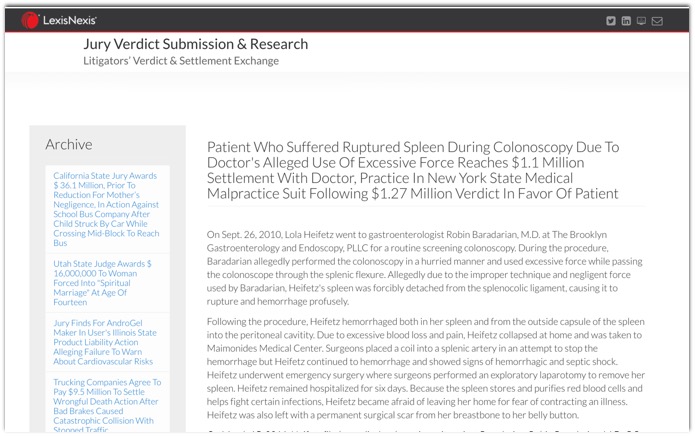
- However, a colonoscopy is the most common cause of iatrogenic splenic injury (in comparison to other procedures or surgeries). The risk factors for splenic injury are both patient and operator dependent. Patient-dependent factors include pre-existing enlargement of the spleen, surgical adhesions, inflammatory bowel disease, and severe diverticular disease. Operator dependent factors include placing the patient on their back, excessive traction, over sedation, slide by advancement, and applying excessive external pressure. Despite these factors, it is still difficult to discern if the complication is unpredictable or directly related to technical factors given rarity of this complication.
- Splenic injury is a rare but serious complication of colonoscopy. Since the mid-1970s, 68 splenic injuries during colonoscopy including our 2 cases have been described. With the increasing use of colonoscopy, endoscopists, surgeons, and radiologists are more likely to encounter this unusual complication. Any cause of increased splenocolic adhesions, splenomegaly, or underlying splenic disease might be a predisposing factor for splenic injury during colonoscopy. However, it can occur in patients without significant adhesions or underlying splenic pathology. The diagnosis is often described in the literature as delayed, because many physicians are not aware of this complication of colonoscopy. Although computerized tomography is highly sensitive, knowledge of this complication is the best tool to aid in early diagnosis. Patients with abdominal pain, hypotension, and a drop in hematocrit without rectal bleeding after colonoscopy should be suspected of having splenic injury.
Splenic injury after elective colonoscopy.
Sarhan M, Ramcharan A, Ponnapalli S.
JSLS. 2009;13(4):616-619.
- However, a colonoscopy is the most common cause of iatrogenic splenic injury (in comparison to other procedures or surgeries). The risk factors for splenic injury are both patient and operator dependent. Patient-dependent factors include pre-existing enlargement of the spleen, surgical adhesions, inflammatory bowel disease, and severe diverticular disease. Operator dependent factors include placing the patient on their back, excessive traction, over sedation, slide by advancement, and applying excessive external pressure. Despite these factors, it is still difficult to discern if the complication is unpredictable or directly related to technical factors given rarity of this complication.
- Splenic injury is a rare but serious complication of colonoscopy. Since the mid-1970s, 68 splenic injuries during colonoscopy including our 2 cases have been described. With the increasing use of colonoscopy, endoscopists, surgeons, and radiologists are more likely to encounter this unusual complication. Any cause of increased splenocolic adhesions, splenomegaly, or underlying splenic disease might be a predisposing factor for splenic injury during colonoscopy. However, it can occur in patients without significant adhesions or underlying splenic pathology. The diagnosis is often described in the literature as delayed, because many physicians are not aware of this complication of colonoscopy. Although computerized tomography is highly sensitive, knowledge of this complication is the best tool to aid in early diagnosis. Patients with abdominal pain, hypotension, and a drop in hematocrit without rectal bleeding after colonoscopy should be suspected of having splenic injury.
Splenic injury after elective colonoscopy.
Sarhan M, Ramcharan A, Ponnapalli S.
JSLS. 2009;13(4):616-619. - Splenic injury is a rare but serious complication of colonoscopy. Since the mid-1970s, 68 splenic injuries during colonoscopy including our 2 cases have been described. With the increasing use of colonoscopy, endoscopists, surgeons, and radiologists are more likely to encounter this unusual complication. Any cause of increased splenocolic adhesions, splenomegaly, or underlying splenic disease might be a predisposing factor for splenic injury during colonoscopy. However, it can occur in patients without significant adhesions or underlying splenic pathology. The diagnosis is often described in the literature as delayed, because many physicians are not aware of this complication of colonoscopy.
Splenic injury after elective colonoscopy.
Sarhan M, Ramcharan A, Ponnapalli S.
JSLS. 2009;13(4):616-619. - The diagnosis is often described in the literature as delayed, because many physicians are not aware of this complication of colonoscopy. Although computerized tomography is highly sensitive, knowledge of this complication is the best tool to aid in early diagnosis. Patients with abdominal pain, hypotension, and a drop in hematocrit without rectal bleeding after colonoscopy should be suspected of having splenic injury.
Splenic injury after elective colonoscopy.
Sarhan M, Ramcharan A, Ponnapalli S.
JSLS. 2009;13(4):616-619. 
- “ Splenic metastases can occur with widespread disease, and parenchymal disease is caused by hematogenous dissemination. The most common primary cancers with splenic metastases include melanoma and cancers of the breast, lung, ovary, stomach, and prostate.”
Nonneoplastic, Benign, and Malignant Splenic Diseases: Cross-Sectional Imaging Findings and Rare Disease Entities Thipphavong S et al. AJR 2014;203: 315-322 - “Lymphoma is the most common malig- nant tumor of the spleen. Lymphoma either can be primary splenic or can be involved in diffuse systemic disease. Splenic involvement is seen at presentation in 33% of all patients with Hodgkin lymphoma and in 30–40% of patients with non-Hodgkin lymphoma.”
Nonneoplastic, Benign, and Malignant Splenic Diseases: Cross-Sectional Imaging Findings and Rare Disease Entities Thipphavong S et al. AJR 2014;203: 315-322 - “Lymphoma can infiltrate the spleen diffusely, causing splenomegaly, or can present as discrete nodules or masses. Necrosis of lymphoma is rare. Infarction of the spleen involved by lymphoma can occur. On ul- trasound, discrete lesions are usually hypoechoic and on CT, lesions are low attenuation, which are best seen on portal venous phase images.”
Nonneoplastic, Benign, and Malignant Splenic Diseases: Cross-Sectional Imaging Findings and Rare Disease Entities Thipphavong S et al. AJR 2014;203: 315-322 - “Splenic angiosarcoma is the most common (nonhematolymphoid) primary malignant neo- plasm of the spleen and arises from the endo- thelial lining of splenic blood vessels. Angio- sarcoma either appears as a well-defined mass or can be diffusely infiltrative in appearance.”
Nonneoplastic, Benign, and Malignant Splenic Diseases: Cross-Sectional Imaging Findings and Rare Disease Entities Thipphavong S et al. AJR 2014;203: 315-322 - “Unlike primary hepatic angiosarcoma, there is no reported association between splenic angiosarcoma and chemical exposures of vinyl chloride, arsenic, or prior injection of Thorotrast, a suspension containing particles of the radioactive compound thorium dioxide that was once used as a radiographic contrast agent. Patients typically present with left upper quadrant pain, anemia, or thrombocytopenia.”
Nonneoplastic, Benign, and Malignant Splenic Diseases: Cross-Sectional Imaging Findings and Rare Disease Entities Thipphavong S et al. AJR 2014;203: 315-322 - “CT shows an enlarged spleen with areas of low and high attenuation, due to acute hemor- rhage or hemosiderin deposits, and calci- fications can be seen. Contrast enhancement of angiosarcoma is variable depending on the degree of tumoral necrosis and can mimic that of a hepatic hemangio- ma by showing avid peripheral enhancement.”
Nonneoplastic, Benign, and Malignant Splenic Diseases: Cross-Sectional Imaging Findings and Rare Disease Entities Thipphavong S et al. AJR 2014;203: 315-322
- Do you need arterial phase imaging for Abdominal Trauma imaging-
- Vascular injury
- Splenic injury
- Hepatic Injury
- Renal Injury - “ The arterial phase of image acquisition improves detection of traumatic contained splenic vascular injuries and should be considered to optimize detection of splenic injuries in trauma with CT.”
Active Hemorrhage and Vascular Injuries in Splenic Trauma: Utility of the Arterial Phase in Multidetector CT
Uyeda JW et al.
Radiology 2014; 270:99-106 - “ More than half of the contained vascular injuries (13 of 22(59%)) were visualized only at the arterial phase of CT, demonstrating that sensitivity and accuracy are improved with the addition of the arterial phase of image acquisition as part of a multiphasic whole body trauma CT.”
Active Hemorrhage and Vascular Injuries in Splenic Trauma: Utility of the Arterial Phase in Multidetector CT
Uyeda JW et al.
Radiology 2014; 270:99-106 - “ The identification of contained vascular injuries is essential because these injuries have an unfavorable outcome with conservative management, with an increased incidence of overt bleeding necessitating blood transfusions, splenectomy with associated lifelong risk of infection, and delayed diagnosis and treatment, potentially leading to increased mortality.”
Active Hemorrhage and Vascular Injuries in Splenic Trauma: Utility of the Arterial Phase in Multidetector CT
Uyeda JW et al.
Radiology 2014; 270:99-106
- “ For CT examination of blunt splenic injury, arterial phase is superior to portal venous phase imaging for pseudoaneurysm but inferior for active bleeding and parenchymal disruption; dual-phase CT provides optimal overall performance.”
Optimizing Trauma Multidetector CT Protocol for Blunt Splenic Injury: Need for Arterial and Portal Venous Phase Scans
Boscak AR et al.
Radiology 2013; 208:79-88 - “ Dual (arterial and portal venous) phase CT has better overall diagnostic performance than single (arterial or portal venous) phase CT for detection of splenic injuries after blunt trauma.”
Optimizing Trauma Multidetector CT Protocol for Blunt Splenic Injury: Need for Arterial and Portal Venous Phase Scans
Boscak AR et al.
Radiology 2013; 208:79-88
