Imaging Pearls ❯ Kidney ❯ Retriperitoneal Masses
|
-- OR -- |
|
- “The perirenal space is a retroperitoneal space that is limited anteriorly by the anterior renal fascia (Gerota fascia) and posteriorly by the posterior renal fascia (Zuckerkandl fascia).These two fasciae fuse to form the lateroconal fascia laterally and blend loosely with the periureteric connective tissue medially. Superiorly, the two fasciae are fixed to the diaphragmatic fascia above the adrenal glands; inferiorly, they blend with the iliac fascia.The anterior and posterior renal fasciae enclose a gradually tapering conelike space produced by the embryologic ascent of the kidneys from the pelvis to the adult retroperitoneal position.”
Neoplastic and Non- neoplastic Proliferative Disorders of the Peri-renal Space: Cross- sectional Imaging Findings
Surabhi VR et al.
RadioGraphics 2008; 28:1005–1017 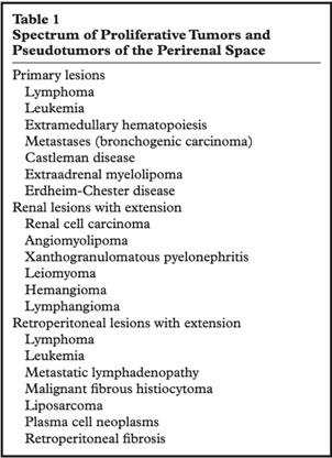
Neoplastic and Non- neoplastic Proliferative Disorders of the Peri-renal Space: Cross- sectional Imaging Findings
Surabhi VR et al.
RadioGraphics 2008; 28:1005–1017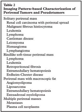
Neoplastic and Non- neoplastic Proliferative Disorders of the Peri-renal Space: Cross- sectional Imaging Findings
Surabhi VR et al.
RadioGraphics 2008; 28:1005–1017- “The perinephric space, shaped as an inverted cone, sits between the anterior and posterior renal fasciae. It can play host to a variety of clinical conditions encountered daily in the reporting schedule for a radiologist. Lesions may be classified and diagnosed based on their imaging characteristics, location and distribution. A broad range of differential diagnoses can be attributed to pathology sitting within this space, often without clinical signs or symptoms. An understanding of commonly encountered conditions affecting the perinephric space, along with characteristic imaging findings, can illustrate and often narrow the likely diagnosis.”
Radiological diagnosis of perinephric pathology: pictorial essay 2015
Goran Mitreski, Tom Sutherland
Insights Imaging (2017) 8:155–169
- Solid Retroperitoneal Tumors: Differential Dx
-Mesodermal neoplasms
-Neurogenic neoplasms
-Germ cell, sex cord, and stromal tumors
-Lymphoid and hematologic neoplasms - Mesodermal Neoplasms: Facts
-Liposarcoma, leiomyosarcoma and MFH make up more than 80% of these tumors
-Most common in 5th and 6th decades of life
-Often aggressive these tumors may be difficult to result and often recur locally or with metastases to liver, lung, bone, and brain - Liposarcoma of the Retroperitoneum: Facts
-Most common retroperitoneal sarcoma (33%)
-10-15% of all liposarcomas occur in the retroperitoneum
-Age range is usually 50-70 years of age
-Usually large with average size of 20 cm
-Four subtypes are well-differentiated, myxoid, pleomorphic and round cell. Well differentiated is most common in retroperitoneum - Retroperitoneal Leiomyosarcoma: facts
-Usually large (>10 cm) when discovered
-More common in woman and usually seen in the 5th to 6th decade of life
-On CT they are usually solid masses but may be cystic and necrotic as well
-6% of Leiomyosarcomas arise from the IVC and are primary in the IVC - Malignant Fibrous Histiocytoma (MFH) of the Retroperitoneum: Facts
-MFH is the most common sarcoma in the body and 15% arise in the retroperitoneum
-More common in male by 3-1 and most common in the 50-60 age group
-Mass may be solid or necrotic and may contain calcification in 7-20% of cases. This may be critical for the differential dx. - Other Retroperitoneal Sarcomas: Facts
-Rhabdomyosarcoma common in younger patients with bimodal peak at age 7 and then teen years
-Angiosarcoma, chondrosarcoma, synovial cell sarcoma all may occur but are rare. Synovial cell sarcoma usually occurs in patient 15-40 years of age which is younger than the other tumors