Imaging Pearls ❯ Chest ❯ Pulmonary Embolism
|
-- OR -- |
|
- Purpose: To evaluate the diagnostic efficacy of artificial intelligence (AI) software in detecting incidental pulmonary embolism (IPE) at CT and shorten the time to diagnosis with use of radiologist reading worklist prioritization.
Conclusion: AI-assisted workflow prioritization of IPE on routine CT scans in oncology patients showed high diagnostic accuracy and significantly shortened the time to diagnosis in a setting with a backlog of examinations.
Artificial Intelligence Tool for Detection and Worklist Prioritization Reduces Time to Diagnosis of Incidental Pulmonary Embolism at CT
Laurens Topff et al.
Radiology: Cardiothoracic Imaging 2023; 5(2):e220163 - Results: In total, 11 736 CT scans in 6447 oncology patients (mean age, 63 years ± 12 [SD]; 3367 men) were included. Prevalence of IPE was 1.3% (51 of 3837 scans), 1.4% (54 of 3920 scans), and 1.0% (38 of 3979 scans) for the respective time periods. The AI software detected 131 true-positive, 12 false-negative, 31 false-positive, and 11 559 true-negative results, achieving 91.6% sensitivity, 99.7% specificity, 99.9% negative predictive value, and 80.9% positive predictive value. During prospective evaluation, AI-based work list prioritization reduced the median DNT for IPE-positive examinations to 87 minutes (vs routine workflow of 7714 minutes and human triage of 4973 minutes). Radiologists’ missed rate of IPE was significantly reduced from 44.8% (47 of 105 scans) without AI to 2.6% (one of 38 scans) when assisted by the AI tool (P < .001).
Artificial Intelligence Tool for Detection and Worklist Prioritization Reduces Time to Diagnosis of Incidental Pulmonary Embolism at CT
Laurens Topff et al.
Radiology: Cardiothoracic Imaging 2023; 5(2):e220163 - “Consequently, long report turnaround times (TATs) pose a risk for delayed diagnosis of unsuspected critical findings. Artificial intelligence (AI) applications that can automatically detect critical findings at imaging can be used to prioritize scans in the reading worklist of the radiologist, with the aim of shortening time to diagnosis and communication with the treating physician. AI-based prioritization tools have been studied for use cases such as intracranial hemorrhage at CT, acute pathologic abnormalities on chest radiographs, and pulmonary embolism (PE) on dedicated CT pulmonary angiograms (CTPAs), with varying results.”
Artificial Intelligence Tool for Detection and Worklist Prioritization Reduces Time to Diagnosis of Incidental Pulmonary Embolism at CT
Laurens Topff et al.
Radiology: Cardiothoracic Imaging 2023; 5(2):e220163 - Key Points
■ Artificial intelligence (AI) software for detecting incidental pulmonary embolism (IPE) at chest CT in patients with cancer showed high diagnostic accuracy in a large sample of 11 736 scans (sensitivity, 91.6%; specificity, 99.7%; negative predictive value, 99.9%).
■ In a practice with a backlog of unreported examinations, AI-based worklist prioritization reduced the median detection and notification time of IPE in flagged scans from several days to 1.0 hour.
■ The missed rate of IPE was significantly reduced from 44.8% to 2.6% when radiologists were assisted by the AI tool (P < .001).
Artificial Intelligence Tool for Detection and Worklist Prioritization Reduces Time to Diagnosis of Incidental Pulmonary Embolism at CT
Laurens Topff et al.
Radiology: Cardiothoracic Imaging 2023; 5(2):e220163 - “The intended use of the investigated AI software is limited to workflow triage and not diagnostics (32). Automated worklist prioritization can assist radiologists, who remain responsible for verifying flagged examinations and must be aware of possible false-negative findings. Our study showed that a considerable number of scans (315 of 4294 [7.3%]) were not analyzed by the deployed AI software due to issues with data retrieval and technical validation, resulting in delayed diagnosis of IPE in these patients. This demonstrates the importance of monitoring and improving the yield of scans analyzed by AI software after deployment.”
Artificial Intelligence Tool for Detection and Worklist Prioritization Reduces Time to Diagnosis of Incidental Pulmonary Embolism at CT
Laurens Topff et al.
Radiology: Cardiothoracic Imaging 2023; 5(2):e220163 - “In conclusion, we demonstrated that commercially available AI software had high diagnostic accuracy in the detection of IPE on chest CT scans in patients with cancer and was effective in significantly reducing the time to diagnosis of positive examinations compared with the routine workflow in a setting with a backlog of unreported scans.”
Artificial Intelligence Tool for Detection and Worklist Prioritization Reduces Time to Diagnosis of Incidental Pulmonary Embolism at CT
Laurens Topff et al.
Radiology: Cardiothoracic Imaging 2023; 5(2):e220163
- Objective: To assess the diagnostic performance of an AI algorithm for detection of iPE on conventional con-trast-enhanced chest CT examinations.
Results: Based on the adjudication process, the frequency of iPE was 1.3% (40/3003). AI detected 4 iPEs missed by clinical reports, and clinical reports detected 7 iPEs missed by AI. AI, compared with clinical reports, exhibited significantly lower specificity (92.7% vs 99.8%, p=.045) and PPV (86.8% vs 97.3%, p=.03), but no significant difference in sensitivity (82.5% vs 90.0%, p=.37) or NPV (99.8% vs 99.9%, p=.36). For AI, neither sensitivity nor specificity varied significantly in association with age, sex, examination location, or cancer-related clinical scenario (all p>.05). Explanations of false positives by AI included metastatic lymph nodes and pulmonary venous filling defect, and of false negatives by AI included surgically altered anatomy and small-caliber subsegmental vessel.
Detection of Incidental Pulmonary Embolism on Conventional Contrast- Enhanced Chest CT: Comparison of an Artificial Intelligence Algorithm and Clinical Reports
Kiran Batra et al.
AJR 2022 Jul 13 [published online]. doi:10.2214/AJR.22.2789 - Results: Based on the adjudication process, the frequency of iPE was 1.3% (40/3003). AI detected 4 iPEs missed by clinical reports, and clinical reports detected 7 iPEs missed by AI. AI, compared with clinical reports, exhibited significantly lower specificity (92.7% vs 99.8%, p=.045) and PPV (86.8% vs 97.3%, p=.03), but no significant differ-ence in sensitivity (82.5% vs 90.0%, p=.37) or NPV (99.8% vs 99.9%, p=.36). For AI, neither sensitivity nor specificity varied significantly in association with age, sex, examination location, or cancer-related clinical scenario (all p>.05). Explanations of false positives by AI included metastatic lymph nodes and pulmonary venous filling defect, and of false negatives by AI included surgically altered anatomy and small-caliber subsegmental vessel.
Conclusion: AI had high NPV and moderate PPV for iPE detection, detecting some iPEs missed by radiologists.
Clinical Impact: Potential applications of the AI tool include serving as a second reader to help detect additional iPEs or as a worklist triage tool to allow earlier iPE detection and intervention. Various explanations of AI misclassifications may provide targets for model improvement.
Detection of Incidental Pulmonary Embolism on Conventional Contrast- Enhanced Chest CT: Comparison of an Artificial Intelligence Algorithm and Clinical Reports
Kiran Batra et al.
AJR 2022 Jul 13 [published online]. doi:10.2214/AJR.22.2789 - KEY FINDING: A commercial AI tool had NPV of 99.8% and PPV of 86.7% for detection of iPE on conventional contrast-enhanced chest CT examinations. Of 40 iPEs present in the study sample, 7 were detected only by the clinical reports, and 4 were detected only by AI.
IMPORTANCE: The AI tool has potential to serve as a second reader or as a worklist triage tool, to facilitate reliable and rapid iPE detection.
Detection of Incidental Pulmonary Embolism on Conventional Contrast- Enhanced Chest CT: Comparison of an Artificial Intelligence Algorithm and Clinical Reports
Kiran Batra et al.
AJR 2022 Jul 13 [published online]. doi:10.2214/AJR.22.2789 - “In conclusion, a commercial AI tool had 99.8% NPV and 86.8% PPV for detection of iPE on conventional contrast-enhanced chest CT examinations (i.e., non-CTPA protocols). Both the AI tool and clinical reports detected iPEs missed by the other method. The diagnostic performance of the AI tool did not show significant variation across study subgroups. Various explanations of misclassifications by the AI tool (both false positives and false negatives) were identified, to provide targets for model improvement. Potential clinical applications of the AI tool include serving as a second reader to help detect additional iPEs missed by radiologists or as a worklist triage and prioritization tool to allow earlier iPE detection and intervention.”
Detection of Incidental Pulmonary Embolism on Conventional Contrast- Enhanced Chest CT: Comparison of an Artificial Intelligence Algorithm and Clinical Reports
Kiran Batra et al.
AJR 2022 Jul 13 [published online]. doi:10.2214/AJR.22.2789 - “The AI algorithm accurately detected 33 of 40 iPEs, including PEs at segmental and subsegmental levels. The AI algorithm identified 4 PEs that were missed by the interpreting radiologists, including by both fellowship-trained cardiothoracic and general radiologists. The findings indicate a potential role to use AI in a second-reader model, to reduce the number of missed PEs. By such an approach, additional potential iPEs identified by AI would undergo dedicated review by the radiologist, who would determine whether the AI-identified finding represents a true iPE that warrants reporting. A tradeoff of this approach is the additional time required for the radiologist to review the AI results, particularly given the numerous false-positive AI findings. Indeed, AI tools in various contexts, including detection of breast cancer, pulmonary nodules, and intracranial hemorrhage, have reported challenges in terms of AI-identified false positives. Thus, optimization of AI algorithms to reducefalse positives will be important to limit fatigue by radiologists in reviewing the AI findings.”
Detection of Incidental Pulmonary Embolism on Conventional Contrast- Enhanced Chest CT: Comparison of an Artificial Intelligence Algorithm and Clinical Reports
Kiran Batra et al.
AJR 2022 Jul 13 [published online]. doi:10.2214/AJR.22.2789
- “The true mortality associated with undiagnosed pulmonary embolism is estimated to be less than 5%, but recovery from pulmonary embolism is associated with complications such as bleeding due to anticoagulant treatment, recurrent venous thromboembolism, chronic thromboembolic pulmonary hypertension, and long-term psychological distress. Approximately half the patients who receive a diagnosis of pulmonary embolism have functional and exercise limitations 1 year later (known as post–pulmonary-embolism syndrome), and the health-related quality of life for patients with a history of pulmonary embolism is diminished as compared with that of matched controls. Therefore, the timely diagnosis and expert management of pulmonary embolism are important.”
Pulmonary Embolism
Susan R. Kahn, Kerstin de Wit
N Engl J Med 2022;387:45-57. 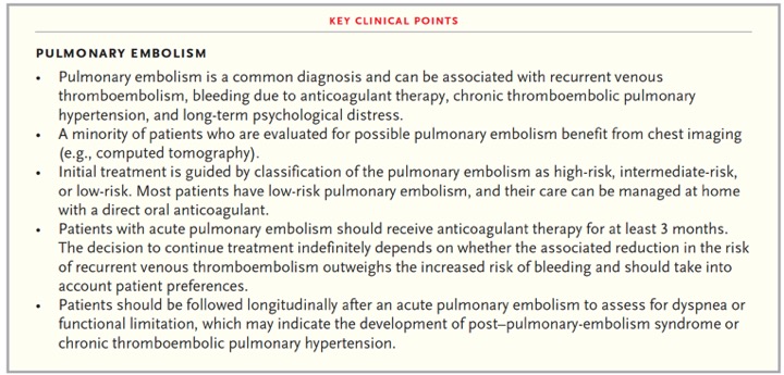
Pulmonary Embolism
Susan R. Kahn, Kerstin de Wit
N Engl J Med 2022;387:45-57.- • Pulmonary embolism is a common diagnosis and can be associated with recurrent venous thromboembolism, bleeding due to anticoagulant therapy, chronic thromboembolic pulmonary hypertension, and long-term psychological distress.
• A minority of patients who are evaluated for possible pulmonary embolism benefit from chest imaging (e.g., computed tomography).
• Initial treatment is guided by classification of the pulmonary embolism as high-risk, intermediate-risk, or low-risk. Most patients have low-risk pulmonary embolism, and their care can be managed at home with a direct oral anticoagulant.
Pulmonary Embolism
Susan R. Kahn, Kerstin de Wit
N Engl J Med 2022;387:45-57. - • Patients with acute pulmonary embolism should receive anticoagulant therapy for at least 3 months. The decision to continue treatment indefinitely depends on whether the associated reduction in the risk of recurrent venous thromboembolism outweighs the increased risk of bleeding and should take into account patient preferences.
• Patients should be followed longitudinally after an acute pulmonary embolism to assess for dyspnea or functional limitation, which may indicate the development of post–pulmonary-embolism syndrome or chronic thromboembolic pulmonary hypertension.
Pulmonary Embolism
Susan R. Kahn, Kerstin de Wit
N Engl J Med 2022;387:45-57. - “Occult cancer is detected in 5.2% of patients within 1 year after a diagnosis of unprovoked pulmonary embolism. An extensive screening strategy may detect more cancers than limited screening, but data are limited as to whether such screening is associated with better patient outcomes. Experts recommend limited cancer screening guided by medical history, physical examination, basic laboratory tests and chest radiographs, and age-specific and sex-specific cancer screening.”
Pulmonary Embolism
Susan R. Kahn, Kerstin de Wit
N Engl J Med 2022;387:45-57. - "Appropriate management of subsegmental pulmonary embolism (a single isolated subsegmental pulmonary embolus or multiple emboli, without the presence of pulmonary embolism in segmental or more proximal pulmonary vessels and without deep-vein thrombosis in the legs) is uncertain. Although some guidelines suggest clinical surveillance instead of anticoagulation in patients with low-risk subsegmental pulmonary embolism, a recent prospective cohort study involving such patients who were treated without anticoagulation therapy showed a higher-than expected incidence of recurrent venous thromboembolism during 90-day follow-up A randomized, placebo-controlled trial of clinical surveillance as compared with anticoagulation in this patient population is ongoing (ClinicalTrials.gov number, NCT04263038).”
Pulmonary Embolism
Susan R. Kahn, Kerstin de Wit
N Engl J Med 2022;387:45-57.

- "Pregnancy is a hypercoagulable state, and consequently pregnant patients are at high risk for PE. The diagnosis of PE can be challenging in pregnant patients, as the clinical signs and symptoms of PE can mimic the physiological effects of pregnancy. In the ATS/STR guidelines, CTPA is the recommended when a chest radiograph is abnormal and therefore maintains an important role in the evaluation of PE. While both V/Q scintigraphy and CTPA have high sensitivities and specificities, CTPA is quick to perform and interpret, and provides a higher rate of alternate diagnoses."
Low dose computed tomography pulmonary angiography protocol for imaging pregnant patients: Can dose reduction be achieved without reducing image quality?
Halpenny D et al.
Clinical Imaging: Volume 44, July–August 2017, Pages 101-105 - "CTPA image quality is potentially impacted by the hemodynamic changes of pregnancy. Increased plasma volume, cardiac output, total vascular resistance, and heart rate can lead to hemodilution of injected intravenous contrast with decreased peak arterial enhancement and shorter contrast material arrival time in the pulmonary arteries. Additionally, the gravid uterus increases IVC pressure and can accentuate transient contrast interruption."
Low dose computed tomography pulmonary angiography protocol for imaging pregnant patients: Can dose reduction be achieved without reducing image quality?
Halpenny D et al.
Clinical Imaging: Volume 44, July–August 2017, Pages 101-105
- OBJECTIVE. The purpose of this study was to assess the difference between mechanical versus hand administration of IV contrast agents on the diagnostic quality of pediatric pulmonary CT angiography (CTA).
CONCLUSION. It is possible to perform diagnostic-quality pulmonary CTA for the assessment of the central pulmonary arteries with hand administration of IV contrast material in pediatric patients with small-gauge IV catheters.
Comparison of Mechanical Versus Hand Administration of IV Contrast Agents for Pediatric Pulmonary CT Angiography Zapala MA et al. AJR 2017; 208:632–636 - “For pediatric patients, pulmonary CTA has become a mainstay for a variety of indications, including pulmonary embolism, pulmonary sling, pulmonary artery hypertension, and pre-or postoperative lung transplants. Pulmonary CTA offers advantages over traditional catheter-based angiography and nuclear medicine ventilation-perfusion studies in terms of improved speed, wider availability, increased contrast and spatial resolution, postprocessing abili- ties, and assessment of other structures outside of the vasculature.”
Comparison of Mechanical Versus Hand Administration of IV Contrast Agents for Pediatric Pulmonary CT Angiography Zapala MA et al. AJR 2017; 208:632–636 - “In conclusion, hand administration of IV contrast material in young children (≤ 5 years) and infants when used in conjunction with a bolus-tracking method yields diagnostic- quality pulmonary CTA studies. This study supports practicing radiologists in the use of hand administration of IV contrast material for pulmonary CTA studies when young children have IV access locations and sizes that preclude mechanical administration.”
Comparison of Mechanical Versus Hand Administration of IV Contrast Agents for Pediatric Pulmonary CT Angiography Zapala MA et al. AJR 2017; 208:632–636 - “The qualities of pulmonary CTA make it desirable to clinicians as a test for suspected PE. The apparent binary yes or no answer to a challenging diagnosis with ambiguous clinical presentation is enticing. As with other diagnostic tests, however, the posttest probability of pulmonary CTA hinges on pretest clinical assessment. Validated clinical decision rules and integrated CDS systems can influence the appropriate use of pulmonary CTA, but further investigation is required to de ne the most successful means of integration into clinical practice.”
Role of Clinical Decision Tools in the Diagnosis of Pulmonary Embolism William M. Sherk, Jadranka Stojanovska AJR 2017; 208:60–70 - “Single or limited educational interventions to inform clinicians of evidence-based decision rules do not overcome the obstacles. Repetitive educational efforts over time—possibly as long as 5 years—in- uence change. Computerized forms of CDS facilitate tracking of individual clinician use and yield of pulmonary CTA and in turn al- low opportunities for feedback. Feedback systems integrated into the electronic medical record show potential for promoting adherence to evidence-based guidelines and use of clinical decision rules before ordering of pulmonary CTA .”
Role of Clinical Decision Tools in the Diagnosis of Pulmonary Embolism William M. Sherk, Jadranka Stojanovska AJR 2017; 208:60–70
- “The incidence of PE is highest in the inpatient setting. While the incidence of PE in outpatient and emergency department patients is similar, more CTA thorax examinations are ordered in the emergency department than in the outpatient setting. There is no difference in the incidence of PE based on who orders CTA thorax examinations.”
Incidence of pulmonary emboli on chest computed tomography angiography based upon referral patterns Meesa IR et al. Emerg Radiol (2016) 23::251–254 - “Our study shows that there is no statistical difference in the incidence of PE when the studies are ordered by attending physicians, residents, or physician extenders. The reasoning might be that in many training institutions, including ours, the attending physician often supervises the residents and physician extenders and, therefore, is often involved in the decision-making process of whether or not to order a CTA to exclude PE. We are somewhat limited in making this conclusion because there are no similar studies in the literature to compare our results against or a way to confirm using the EMR and a retrospective methodology.”
Incidence of pulmonary emboli on chest computed tomography angiography based upon referral patterns Meesa IR et al. Emerg Radiol (2016) 23::251–254
- "Practical measures to reduce the risk of PE misdiagnosis could and should include any of the following: systematic use of pretest probability assessment (which would require buy-in from clinicians and incorporation into imaging protocols); radiology technologists being educated to optimize image quality, focusing on proper patient breathing technique and repeating examinations where appropriate; increased familiarization by radiologists with the range of potential diag- nostic pitfalls; encouragement of the use of second opinions by interpreting radiologists, particularly for solitary subsegmental PEs; and regular review of positive pulmonary CTA cases (e.g., at monthly discrepancy or audit meetings). Some of these measures are easier to implement than others, but their importance is underscored by the implications of a false-positive diagnosis of PE.”
Overdiagnosis of Pulmonary Embolism by Pulmonary CT Angiography Hutchinson BD et al. AJR 2015; 205:271–277 - "Practical measures to reduce the risk of PE misdiagnosis could and should include any of the following: systematic use of pretest probability assessment (which would require buy-in from clinicians and incorporation into imaging protocols); radiology technologists being educated to optimize image quality, focusing on proper patient breathing technique and repeating examinations where appropriate; increased familiarization by radiologists with the range of potential diag- nostic pitfalls; encouragement of the use of second opinions by interpreting radiologists, particularly for solitary subsegmental PEs; and regular review of positive pulmonary CTA cases (e.g., at monthly discrepancy or audit meetings).”
Overdiagnosis of Pulmonary Embolism by Pulmonary CT Angiography Hutchinson BD et al. AJR 2015; 205:271–277 - “In routine clinical practice, PEs diagnosed by pulmonary CTA are frequently overdiagnosed, when compared with the consensus opinion of a panel of expert chest radiologists. Improvements in the quality of pulmonary CTA examination and increased fa- miliarity with potential diagnostic pitfalls in pulmonary CTA are recommended to minimize misdiagnosis of PE.”
Overdiagnosis of Pulmonary Embolism by Pulmonary CT Angiography Hutchinson BD et al. AJR 2015; 205:271–277 - “A total of 937 pulmonary CTA studies were performed over the study period. PE was diagnosed in the initial report in 174 of these cases (18.6%). There was discordance between the chest radiologists and the original radiologist in 45 of 174 (25.9%) cases. Discordance occurred more often where the original reported PE was solitary (46.2% of reported solitary PEs were considered negative on retrospective review) and located in a segmental or subsegmental pulmonary artery (26.8% of segmental and 59.4% of subsegmental PE diagnoses were considered negative on retrospective review). The most common cause of diagnostic
difficulty was breathing motion artifact, followed by beam-hardening artifact.”
Overdiagnosis of Pulmonary Embolism by Pulmonary CT Angiography Hutchinson BD et al. AJR 2015; 205:271–277 - “The risk of hemorrhage related to anticoagulation therapy is potentially significant. A large meta-analysis in 2003 [8] found a 7% annual risk of major bleeding and a 0.4% incidence of bleeding-related fatality in pa- tients treated with oral anticoagulation therapy for venous thromboembolism for longer than 3 months. The practical implications of long-term anticoagulation therapy for the patient are also potentially significant, requir- ing frequent attendance to their medical practitioners for blood tests, consequent time off from work, potential adverse drug interactions with other medications, adjustments to travel and lifestyle, implications for future dental and medical procedures, and possible negative effects on life insurance status.”
Overdiagnosis of Pulmonary Embolism by Pulmonary CT Angiography Hutchinson BD et al. AJR 2015; 205:271–277 - “This study shows an unexpectedly high rate of overdiagnosis of PE by pulmonary CTA in a tertiary-care university hospital, with an overall rate of 25.9% of all positive pulmonary CTA examinations, increasing to as high as 66.7% of cases where a solitary subsegmental PE was originally reported. Discordance was greatest for solitary PEs, PEs located in segmental and subsegmental pulmonary arteries, and in the lower zones of the lungs. The positive predictive value of pulmonary CTA for the diagnosis of PE was only 74.1% in this study.”
Overdiagnosis of Pulmonary Embolism by Pulmonary CT Angiography Hutchinson BD et al. AJR 2015; 205:271–277 

- “ This study shows that missed PE can occur on abdominal CT. It is recommended that interpretation include a careful search of the lower pulmonary arterial vasculature on contrast-enhanced abdominal CT scans.”
Missed Pulmonary Embolism on Abdominal CT
Lim KY, Kligerman SJ, Lin CT, White CS
AJR 2014; 202:738-743 - “ The challenge in identifying PE is clearly greater on abdominal CT than on chest CT. In addition to the multiple pitfalls described already, the lungs are typically not a primary target of abdominal CT interpretation, and only a limited part of the pulmonary anatomy is included.”
Missed Pulmonary Embolism on Abdominal CT
Lim KY, Kligerman SJ, Lin CT, White CS
AJR 2014; 202:738-743
- “ Septic pulmonary embolism (SPE) is an uncommon disorder that generally presents with an insidious onset of fever, respiratory symptoms, and lung infiltrates. Clinical and radiologic features at presentation are usually nonspecific, and the diagnosis of this disorder is frequently delayed. Historically, SPE has been associated with risk factors such as IV drug use, pelvic thrombophlebitis, and suppurative processes in the head and neck.However, increasing use of indwelling catheters and devices as well as increasing numbers of immunocompromised patients have changed the epidemiology and clinical manifestations of SPE.”
Septic Pulmonary Embolism: Presenting Features and Clinical Course of 14 Patients
Cook RJ et al.
Chest 2005;128(1):162-166 - “Historically, SPE has been associated with risk factors such as IV drug use, pelvic thrombophlebitis, and suppurative processes in the head and neck.However, increasing use of indwelling catheters and devices as well as increasing numbers of immunocompromised patients have changed the epidemiology and clinical manifestations of SPE.”
Septic Pulmonary Embolism: Presenting Features and Clinical Course of 14 Patients
Cook RJ et al.
Chest 2005;128(1):162-166 - “ Septic pulmonary embolism (SPE) is an uncommon disorder that generally presents with an insidious onset of fever, respiratory symptoms, and lung infiltrates. Clinical and radiologic features at presentation are usually nonspecific, and the diagnosis of this disorder is frequently delayed.”
Septic Pulmonary Embolism: Presenting Features and Clinical Course of 14 Patients
Cook RJ et al.
Chest 2005;128(1):162-166 - “Although findings on CXR tend to be nonspecific, CT may yield helpful clues that may suggest the diagnosis of SPE. Parenchymal lesions related to SPE are usually multiple and nodular with a peripheral distribution and a tendency for cavitation. These features associated with an extrapulmonary focus of infection should lead to consideration of SPE as the cause. Although other authors have described a “feeding vessel” sign (a vessel leading to a peripheral lung lesion) as a characteristic feature of SPE, we were able to identify this feature associated with only a minority of parenchymal lesions and did not find it particularly helpful in the recognition of SPE.”
Septic Pulmonary Embolism: Presenting Features and Clinical Course of 14 Patients
Cook RJ et al.
Chest 2005;128(1):162-166