Wrist Pathology: Other Carpal Fractures
Identification of carpal fractures other than the scaphoid can be difficult on conventional radiographs. The second most frequently fractured carpal bone is the triquetrum. Types of fractures include the dorsal cortex, volar cortex and triquetral body. Body fractures usually occur in conjunction with other carpal fractures and ligamentous injury to the wrist. The fracture of the dorsal cortex can be identified as an avulsion fragment on the lateral radiograph. This type of fracture usually follows a fall on the dorsiflexed wrist or avulsion by the radiotriquetral and scaphotriquetral ligaments, which insert on the dorsal surface of the triquetral bone. Avulsion fractures can also arise from the volar surface of the bone. This type of fracture is not readily identified on the three radiographic views of the wrist, but requires an instability series, CT or MR to detect. Unfortunately. this subtle fracture is associated with injury to the perilunate ligaments resulting in lunatotriquetral instability and long term pain and carpal dysfunction. If this type of fracture is identified, correlative imaging should be performed with MR respectively, to evaluate the perilunate ligaments.
Fractures of the hamate most commonly involve the hamate hook. This type of injury is usually caused by direct trauma by swinging the handle of a baseball bat, golf club or tennis racquet, but can infrequently result from trauma directed to the proximal palm. Traditional radiographic views of the wrist are usually insufficient to identify this fracture. Carpal tunnel radiographs or supinated oblique views of the wrist are required; however, even these views may not demonstrate the fracture, necessitating CT. CT in the axial plane is best for evaluation of the hamate hook. Sagittal displays are better for the body of the hamate. Diagnosis is important because the ulnar nerve travels near the hamate hook, and neurologic damage can result from the motion of an ununited fracture.
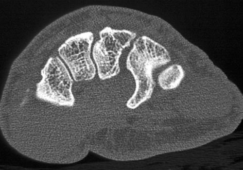 |  |  |  |
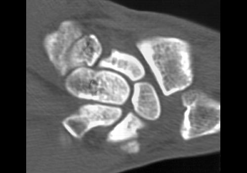
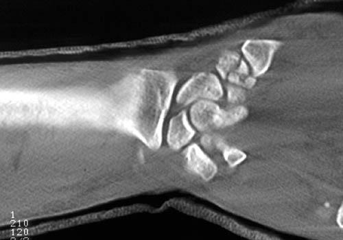
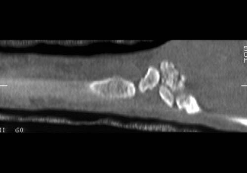
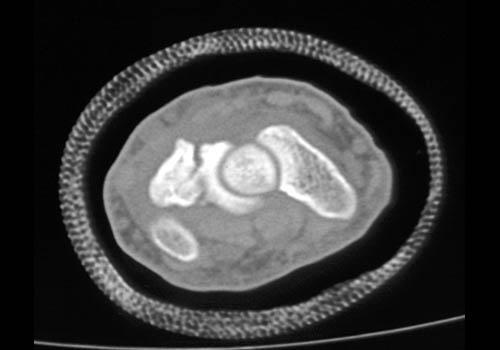
Clinical History: 50 year old man with history of major trauma to both hands and arms years ago. He has persistant pain. CT Findings: A tiny area of ossification is identified adjacent to the lunate, well circumscribed with smooth margins. No evidence of osteomyelitis. Diagnosis: Bone fragment adjacent to lunate due to previous trauma. Outcome: Comments: | |||
 | 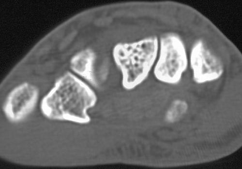 |  |  |
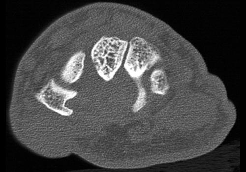
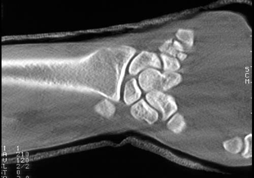
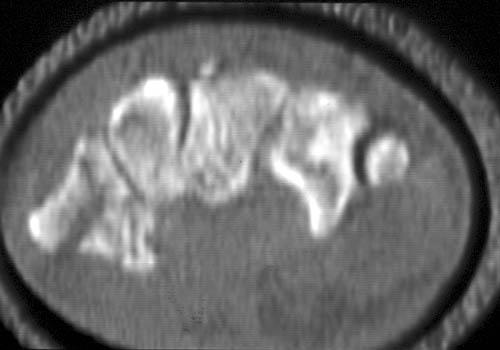
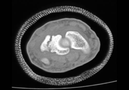
Clinical History This patient had been experiencing continued pain. CT Findings: CT scan demonstrates a hamate fracture. Diagnosis: Hamate fracture. Outcome: Comments: The fracture lines are fairly smooth and the fracture had happened a while prior to the CT scan. | |||
 |  |  |  |
|  |  |  |
 | |||
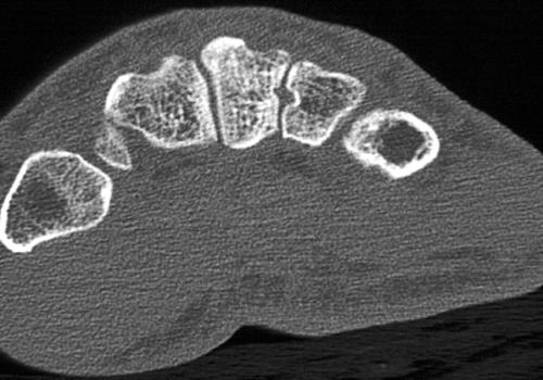
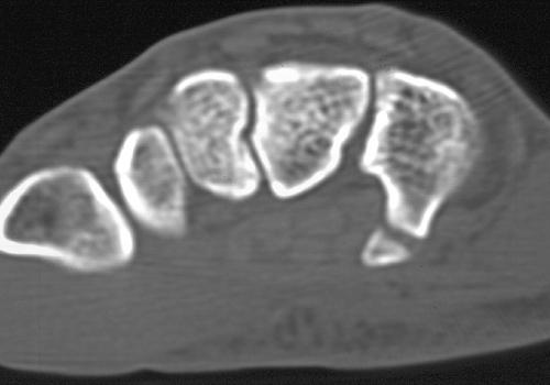
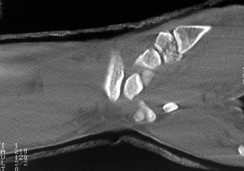
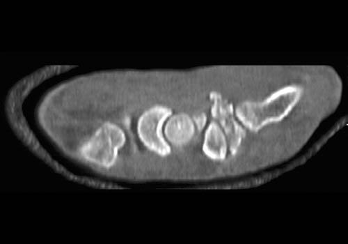
Clinical History: 28 year old man. CT Findings: Comminuted fracture of the right pisiform. Diagnosis: Pisiform fracture. Outcome: Comments: | |||
 |  |  | |
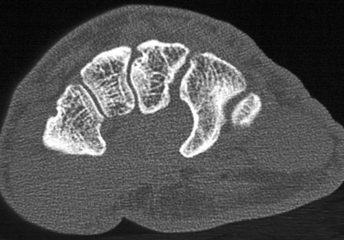
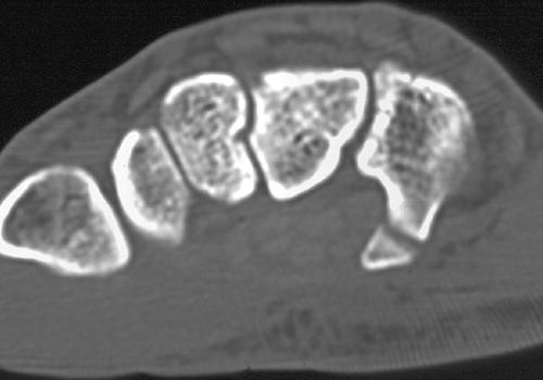
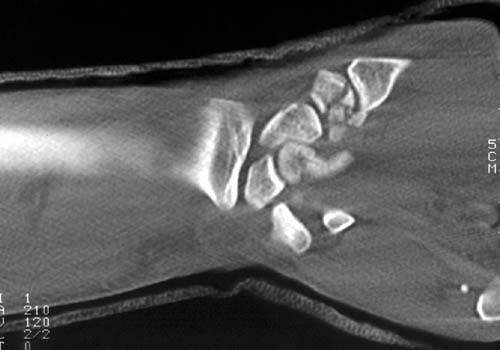
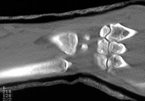
Clinical History: 16 year old boy with lunatotriquetral tear. He underwent triquetral-lunate fusion CT Findings: Diagnosis: Triquetral-lunate fusion Outcome: Comments: | |||